The UBE4A Antibody (CAB3354) is a high-quality antibody developed for reliable detection and analysis of target proteins. This antibody, raised in rabbits, exhibits high reactivity with human samples and is validated for use in Western blot applications. By binding specifically to the UBE4A protein, this antibody enables precise detection and analysis in various cell types, making it ideal for studies in molecular biology and disease mechanisms.UBE4A is known for its function in targeting misfolded proteins for degradation, a process crucial for maintaining cellular homeostasis and preventing the accumulation of damaged proteins.
This antibody is validated for use in WB, IHC-P, ELISA applications and has demonstrated reactivity against Human, Mouse, Rat samples.
Product Name:
UBE4A Antibody
SKU:
CAB3354
Size:
20μL, 100μL
Reactivity:
Human, Mouse, Rat
Conjugate:
Unconjugated
Immunogen:
Recombinant protein (or fragment).This information is considered to be commercially sensitive.
Recommended starting concentration is 1 μg/mL. Please optimize the concentration based on your specific assay requirements.
Synonyms:
E4, UFD2, UBOX2, NEDHMS, UBE4A
Positive Sample:
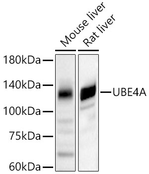
Mouse liver, Rat liver
Cellular Localization:
Cytoplasm.
Calculated MW:
123kDa
Observed MW:
123kDa
This gene encodes a member of the U-box ubiquitin ligase family. The encoded protein is involved in multiubiquitin chain assembly and plays a critical role in chromosome condensation and separation through the polyubiquitination of securin. Autoantibodies against the encoded protein may be markers for scleroderma and Crohn's disease. A pseudogene of this gene is located on the long arm of chromosome 3. Alternatively spliced transcript variants encoding multiple isoforms have been observed for this gene.
Purification Method
Affinity purification
Gene ID
9354
RRID
AB_2765070
Buffer Information
Store at -20℃. Avoid freeze / thaw cycles. Buffer: PBS containing 50% glycerol, preserved with proclin300 or sodium azide, pH 7.3.
Western blot analysis of various lysates, using UBE4A Rabbit pAb (CAB3354) at 1:3000 dilution. Secondary antibody: HRP-conjugated Goat anti-Rabbit IgG (H+L) (CABS014) at 1:10000 dilution. Lysates/proteins: 25μg per lane. Blocking buffer: 3% nonfat dry milk in TBST. Detection: ECL Basic Kit (AbGn00020). Exposure time: 60s.
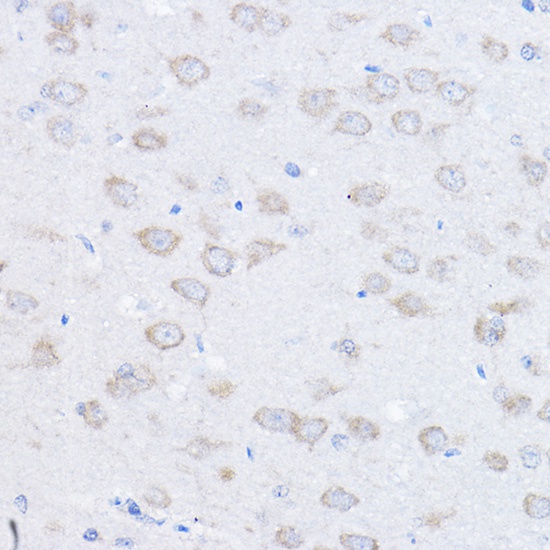
Immunohistochemistry analysis of paraffin-embedded Rat brain using UBE4A Rabbit pAb (CAB3354) at dilution of 1:100 (40x lens). High pressure antigen retrieval performed with 0.01M Citrate buffer (pH 6.0) prior to IHC staining.