The UQCRC1 Antibody (CAB3339) is a high-quality antibody developed for reliable detection and analysis of target proteins. This antibody, raised in rabbits, exhibits high reactivity with human samples and is ideal for use in Western blot applications. By binding to the UQCRC1 protein, this antibody enables researchers to detect and analyze UQCRC1 expression in various cell types, making it useful for investigations in the fields of bioenergetics, mitochondrial function, and diseases related to mitochondrial dysfunction.UQCRC1 is a crucial component of the electron transport chain, playing a vital role in generating ATP, the energy currency of the cell.
This antibody is validated for use in WB, IHC-P, IF/ICC, ELISA applications and has demonstrated reactivity against Human, Mouse, Rat samples.
Product Name:
UQCRC1 Antibody
SKU:
CAB3339
Size:
20μL, 100μL
Reactivity:
Human, Mouse, Rat
Conjugate:
Unconjugated
Immunogen:
Recombinant protein (or fragment).This information is considered to be commercially sensitive.
Recommended starting concentration is 1 μg/mL. Please optimize the concentration based on your specific assay requirements.
Synonyms:
QCR1, PKNPY, UQCR1, D3S3191, UQCRC1
Positive Sample:
HeLa, Hep G2, 293T, NIH/3T3, Rat spleen
Cellular Localization:
Mitochondrion Inner Membrane.
Calculated MW:
53kDa
Observed MW:
53kDa
Enables ubiquitin protein ligase binding activity. Predicted to be involved in oxidative phosphorylation. Predicted to act upstream of or within mitochondrial electron transport, ubiquinol to cytochrome c. Located in mitochondrion. Implicated in Alzheimer's disease. Biomarker of Alzheimer's disease.
Purification Method
Affinity purification
Gene ID
7384
RRID
AB_2765058
Buffer Information
Store at -20℃. Avoid freeze / thaw cycles. Buffer: PBS containing 50% glycerol, preserved with proclin300 or sodium azide, pH 7.3.
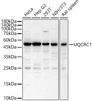
Western blot analysis of various lysates, using UQCRC1 Rabbit pAb (CAB3339) at 1:1000 dilution. Secondary antibody: HRP-conjugated Goat anti-Rabbit IgG (H+L) (CABS014) at 1:10000 dilution. Lysates/proteins: 25μg per lane. Blocking buffer: 3% nonfat dry milk in TBST. Detection: ECL Basic Kit (AbGn00020). Exposure time: 30s.
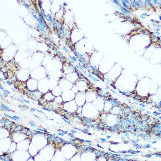
Immunohistochemistry analysis of paraffin-embedded Human colon using UQCRC1 Rabbit pAb (CAB3339) at dilution of 1:100 (40x lens). Microwave antigen retrieval performed with 0.01M Tris/EDTA Buffer (pH 9.0) prior to IHC staining.
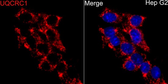
Immunofluorescence analysis of HepG2 cells using UQCRC1 Rabbit pAb (CAB3339) at dilution of 1:100 (40x lens). Secondary antibody: Cy3-conjugated Goat anti-Rabbit IgG (H+L) (CABS007) at 1:500 dilution. Blue: DAPI for nuclear staining.