The UPK3A Antibody (CAB10034) is a high-quality antibody developed for reliable detection and analysis of target proteins. This antibody, produced in rabbits, exhibits high specificity and sensitivity for detecting uroplakin 3A in human samples, making it suitable for Western blot applications.Uroplakin 3A is a key component of urothelial cells, playing a crucial role in maintaining the integrity and barrier function of the bladder epithelium. Dysregulation of uroplakin 3A expression has been implicated in various bladder disorders and diseases, making it a target of interest in urological research.
This antibody is validated for use in WB, ELISA, IF-P applications and has demonstrated reactivity against Human, Mouse, Rat samples.
Product Name:
UPK3A Antibody
SKU:
CAB10034
Size:
20μL, 100μL
Reactivity:
Human, Mouse, Rat
Conjugate:
Unconjugated
Immunogen:
Recombinant protein (or fragment).This information is considered to be commercially sensitive.
Recommended starting concentration is 1 μg/mL. Please optimize the concentration based on your specific assay requirements.
Synonyms:
UP3A, UPK3, UPIII, UPIIIA, UPK3A
Positive Sample:
HeLa, HL-60, Mouse brain, Mouse kidney, Rat kidney
Cellular Localization:
Endoplasmic Reticulum Membrane, Single-Pass Type I Membrane Protein.
Calculated MW:
31kDa
Observed MW:
30kDa
This gene encodes a member of the uroplakin family, a group of transmembrane proteins that form complexes on the apical surface of the bladder epithelium. Mutations in this gene may be associated with renal adysplasia. Alternatively spliced transcript variants have been described.
Purification Method
Affinity purification
Gene ID
7380
RRID
AB_2757555
Buffer Information
Store at -20℃. Avoid freeze / thaw cycles. Buffer: PBS containing 50% glycerol, preserved with proclin300 or sodium azide, pH 7.3.
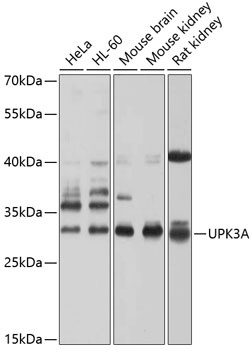
Western blot analysis of various lysates using UPK3A Rabbit pAb (CAB10034) at 1:1000 dilution. Secondary antibody: HRP-conjugated Goat anti-Rabbit IgG (H+L) (CABS014) at 1:10000 dilution. Lysates/proteins: 25μg per lane. Blocking buffer: 3% nonfat dry milk in TBST. Detection: ECL Basic Kit (AbGn00020). Exposure time: 5s.
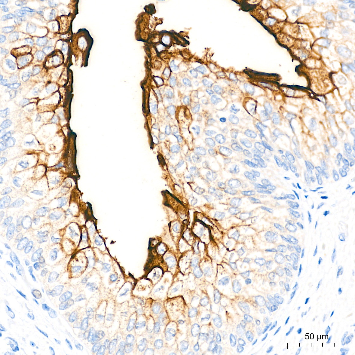
Immunohistochemistry analysis of paraffin-embedded Human bladder tissue using UPK3A Rabbit pAb (CAB10034) at a dilution of 1:4000 (40x lens). High pressure antigen retrieval performed with 0.01M Tris-EDTA Buffer (pH 9.0) prior to IHC staining.
Immunohistochemistry analysis of paraffin-embedded Rat bladder tissue using UPK3A Rabbit pAb (CAB10034) at a dilution of 1:4000 (40x lens). High pressure antigen retrieval performed with 0.01M Tris-EDTA Buffer (pH 9.0) prior to IHC staining.
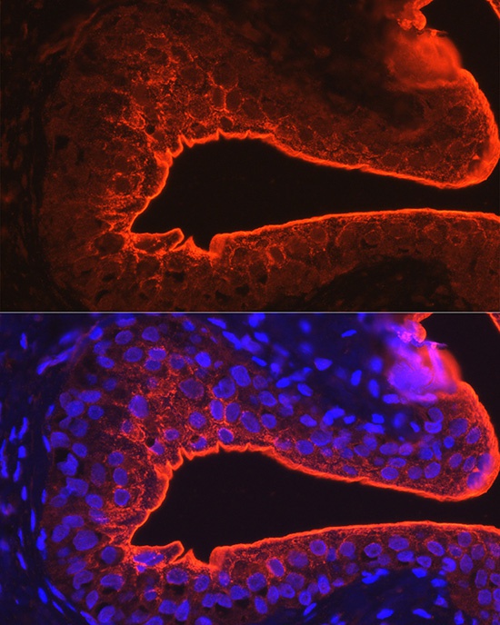
Immunofluorescence analysis of paraffin-embedded rat bladder using UPK3A Rabbit pAb (CAB10034) at dilution of 1:100 (40x lens). Secondary antibody: Cy3-conjugated Goat anti-Rabbit IgG (H+L) (CABS007) at 1:500 dilution. Blue: DAPI for nuclear staining.