The VAMP7 Antibody (CAB18698) is a high-quality antibody developed for reliable detection and analysis of target proteins. This antibody, generated in rabbits, exhibits high reactivity with human samples and has been validated for use in Western blot applications.VAMP7 is known to play a crucial role in regulating intracellular membrane trafficking, particularly within the endosomal-lysosomal pathway. Dysfunction of VAMP7 has been implicated in various diseases, including neurodegenerative disorders and infectious diseases.
This antibody is validated for use in WB, IF/ICC, ELISA applications and has demonstrated reactivity against Human, Mouse, Rat samples.
Product Name:
VAMP7 Antibody
SKU:
CAB18698
Size:
20μL, 100μL
Reactivity:
Human, Mouse, Rat
Immunogen:
Recombinant protein (or fragment).This information is considered to be commercially sensitive.
This gene encodes a transmembrane protein that is a member of the soluble N-ethylmaleimide-sensitive factor attachment protein receptor (SNARE) family. The encoded protein localizes to late endosomes and lysosomes and is involved in the fusion of transport vesicles to their target membranes. Alternate splicing results in multiple transcript variants.
Purification Method
Affinity purification
Gene ID
6845
RRID
AB_2862432
Buffer Information
Store at -20℃. Avoid freeze / thaw cycles. Buffer: PBS containing 50% glycerol, preserved with proclin300 or sodium azide, pH 7.3.
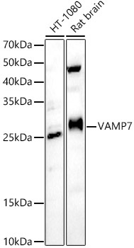
Western blot analysis of various lysates, using VAMP7 Rabbit pAb (CAB18698) at 1:500 dilution. Secondary antibody: HRP-conjugated Goat anti-Rabbit IgG (H+L) (CABS014) at 1:10000 dilution. Lysates/proteins: 25μg per lane. Blocking buffer: 3% nonfat dry milk in TBST. Detection: ECL Enhanced Kit (AbGn00021). Exposure time: 60s.
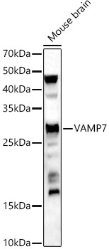
Western blot analysis of lysates from Mouse brain, using VAMP7 Rabbit pAb (CAB18698) at 1:500 dilution. Secondary antibody: HRP-conjugated Goat anti-Rabbit IgG (H+L) (CABS014) at 1:10000 dilution. Lysates/proteins: 25μg per lane. Blocking buffer: 3% nonfat dry milk in TBST. Detection: ECL Enhanced Kit (AbGn00021). Exposure time: 180s.
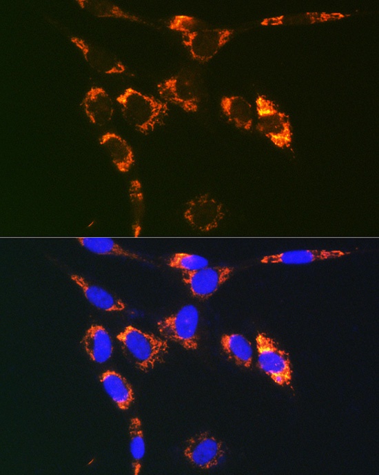
Immunofluorescence analysis of NIH/3T3 cells using VAMP7 Rabbit pAb (CAB18698) at dilution of 1:100. Secondary antibody: Cy3-conjugated Goat anti-Rabbit IgG (H+L) (CABS007) at 1:500 dilution. Blue: DAPI for nuclear staining.
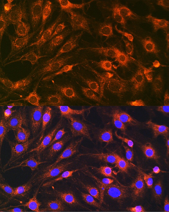
Immunofluorescence analysis of C6 cells using VAMP7 Rabbit pAb (CAB18698) at dilution of 1:100. Secondary antibody: Cy3-conjugated Goat anti-Rabbit IgG (H+L) (CABS007) at 1:500 dilution. Blue: DAPI for nuclear staining.