The VASP Antibody (CAB0166) is a high-quality antibody developed for reliable detection and analysis of target proteins. This antibody, produced in rabbits, exhibits high specificity and reactivity with human samples, making it an ideal choice for Western blot applications. By targeting the VASP protein, researchers can analyze its expression and function in various cell types, providing insights into processes such as cell migration, adhesion, and signaling.VASP, also known as vasodilator-stimulated phosphoprotein, is a key player in actin filament assembly and organization, influencing cell movement and shape changes.
This antibody is validated for use in WB, ELISA applications and has demonstrated reactivity against Human samples.
Product Name:
VASP Antibody
SKU:
CAB0166
Size:
20μL, 100μL
Reactivity:
Human
Conjugate:
Unconjugated
Immunogen:
Synthetic peptide. This information is considered to be commercially sensitive.
Vasodilator-stimulated phosphoprotein (VASP) is a member of the Ena-VASP protein family. Ena-VASP family members contain an EHV1 N-terminal domain that binds proteins containing E/DFPPPPXD/E motifs and targets Ena-VASP proteins to focal adhesions. In the mid-region of the protein, family members have a proline-rich domain that binds SH3 and WW domain-containing proteins. Their C-terminal EVH2 domain mediates tetramerization and binds both G and F actin. VASP is associated with filamentous actin formation and likely plays a widespread role in cell adhesion and motility. VASP may also be involved in the intracellular signaling pathways that regulate integrin-extracellular matrix interactions. VASP is regulated by the cyclic nucleotide-dependent kinases PKA and PKG.
Purification Method
Affinity purification
Gene ID
7408
RRID
AB_2756994
Buffer Information
Store at -20℃. Avoid freeze / thaw cycles. Buffer: PBS containing 50% glycerol, preserved with proclin300 or sodium azide, pH 7.3.
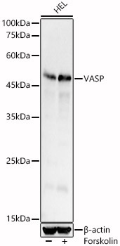
Western blot analysis of lysates from HEL cells, using VASP Rabbit pAb (CAB0166) at 1:600 dilution. HEL cells were treated with Forskolin (10 uM) at 37℃ for 1 hour after serum-starvation overnight. Secondary antibody: HRP-conjugated Goat anti-Rabbit IgG (H+L) (CABS014) at 1:10000 dilution. Lysates/proteins: 25μg per lane. Blocking buffer: 3% nonfat dry milk in TBST. Detection: ECL Basic Kit (AbGn00020). Exposure time: 10s.