The VASP Antibody (CAB14217) is a high-quality antibody developed for reliable detection and analysis of target proteins. This antibody is generated in rabbits and is highly specific to human VASP protein, making it a reliable choice for Western blot applications. By binding to VASP, this antibody allows for the detection and analysis of VASP expression in various cell types, making it ideal for studies in cell biology, cancer metastasis, and cardiovascular research.VASP is a crucial protein involved in cellular processes such as cell adhesion, migration, and invasion, making it a target of interest in cancer progression and metastasis.
This antibody is validated for use in WB, IHC-P, IF/ICC, ELISA applications and has demonstrated reactivity against Human, Mouse, Rat samples.
Product Name:
VASP Antibody
SKU:
CAB14217
Size:
20μL, 100μL
Reactivity:
Human, Mouse, Rat
Conjugate:
Unconjugated
Immunogen:
Synthetic peptide. This information is considered to be commercially sensitive.
Vasodilator-stimulated phosphoprotein (VASP) is a member of the Ena-VASP protein family. Ena-VASP family members contain an EHV1 N-terminal domain that binds proteins containing E/DFPPPPXD/E motifs and targets Ena-VASP proteins to focal adhesions. In the mid-region of the protein, family members have a proline-rich domain that binds SH3 and WW domain-containing proteins. Their C-terminal EVH2 domain mediates tetramerization and binds both G and F actin. VASP is associated with filamentous actin formation and likely plays a widespread role in cell adhesion and motility. VASP may also be involved in the intracellular signaling pathways that regulate integrin-extracellular matrix interactions. VASP is regulated by the cyclic nucleotide-dependent kinases PKA and PKG.
Purification Method
Affinity purification
Gene ID
7408
RRID
AB_2761077
Buffer Information
Store at -20℃. Avoid freeze / thaw cycles. Buffer: PBS containing 50% glycerol, preserved with proclin300 or sodium azide, pH 7.3.
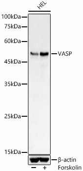
Western blot analysis of lysates from HEL cells, using VASP Rabbit pAb (CAB14217) at 1:800 dilution. HEL cells were treated with Forskolin (10 uM) at 37℃ for 1 hour after serum-starvation overnight. Secondary antibody: HRP-conjugated Goat anti-Rabbit IgG (H+L) (CABS014) at 1:10000 dilution. Lysates/proteins: 25μg per lane. Blocking buffer: 3% nonfat dry milk in TBST. Detection: ECL Basic Kit (AbGn00020). Exposure time: 3s.
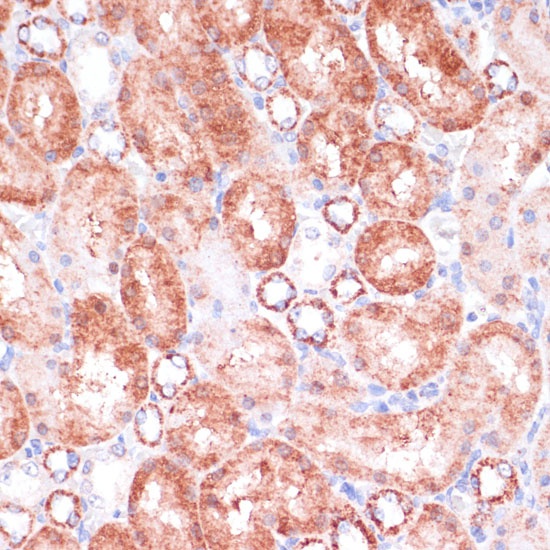
Immunohistochemistry analysis of paraffin-embedded Rat kidney using VASP Rabbit pAb (CAB14217) at dilution of 1:100 (40x lens). Microwave antigen retrieval performed with 0.01M PBS Buffer (pH 7.2) prior to IHC staining.
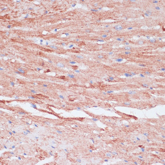
Immunohistochemistry analysis of paraffin-embedded Mouse heart using VASP Rabbit pAb (CAB14217) at dilution of 1:100 (40x lens). Microwave antigen retrieval performed with 0.01M PBS Buffer (pH 7.2) prior to IHC staining.
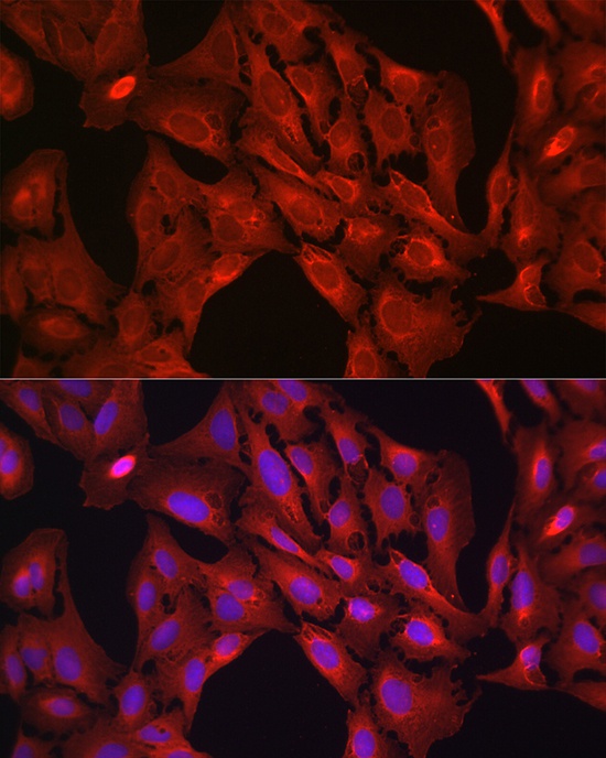
Immunofluorescence analysis of U-2 OS cells using VASP Rabbit pAb (CAB14217) at dilution of 1:100 (40x lens). Secondary antibody: Cy3-conjugated Goat anti-Rabbit IgG (H+L) (CABS007) at 1:500 dilution. Blue: DAPI for nuclear staining.