The VCAM1 Antibody (CAB0279) is a high-quality antibody developed for reliable detection and analysis of target proteins. This gene is a member of the Ig superfamily and encodes a cell surface sialoglycoprotein expressed by cytokine-activated endothelium. This type I membrane protein mediates leukocyte-endothelial cell adhesion and signal transduction, and may play a role in the development of artherosclerosis and rheumatoid arthritis. Three alternatively spliced transcripts encoding different isoforms have been described for this gene. RRID AB_2757091 Gene ID 7412 Swiss Prot Synonym CD106; INCAM-100; VCAM1
This antibody is validated for use in WB, ELISA, IF-P applications and has demonstrated reactivity against Human, Mouse samples.
Product Name:
VCAM1 Antibody
SKU:
CAB0279
Size:
100μL, 20μL
Reactivity:
Human, Mouse
Clone Number:
-
Conjugate:
Unconjugated
Immunogen:
Synthetic peptide. This information is considered to be commercially sensitive.
Tested Applications:
WBELISAIF-P
Recommended Dilution:
WB
1:500 - 1:1000
IF-P
1:50 - 1:200
ELISA
Recommended starting concentration is 1 μg/mL. Please optimize the concentration based on your specific assay requirements.
Synonyms:
CD106, INCAM-100, VCAM1
Positive Sample:
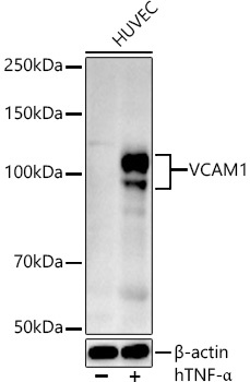
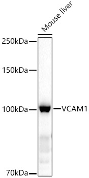
HUVEC treated with hTNF-α, Mouse liver
Cellular Localization:
Membrane, Single-Pass Type I Membrane Protein.
Calculated MW:
81kDa
Observed MW:
95-120kDa
This gene is a member of the Ig superfamily and encodes a cell surface sialoglycoprotein expressed by cytokine-activated endothelium. This type I membrane protein mediates leukocyte-endothelial cell adhesion and signal transduction, and may play a role in the development of artherosclerosis and rheumatoid arthritis. Three alternatively spliced transcripts encoding different isoforms have been described for this gene. RRID AB_2757091 Gene ID 7412 Swiss Prot Synonym CD106; INCAM-100; VCAM1
Purification Method:
Affinity purification
Gene ID:
7412
RRID:
AB_2757091
Buffer Information:
Store at -20℃. Avoid freeze / thaw cycles. Buffer: PBS containing 50% glycerol, preserved with proclin300 or sodium azide, pH 7.3.
Western blot analysis of lysates from HUVEC cells, using VCAM1 Rabbit pAb (CAB0279) at 1:500 dilution. HUVEC cells were treated with Human Tumor Necrosis Factor-α (hTNF-α) (10 ng/ml) for 16 hours. Secondary antibody: HRP-conjugated Goat anti-Rabbit IgG (H+L) (AS014) at 1:10000 dilution. Lysates/proteins: 25μg per lane. Blocking buffer: 3% nonfat dry milk in TBST. Detection: ECL Basic Kit (AbGn00020). Exposure time: 90s.
Western blot analysis of lysates from Mouse liver, using VCAM1 Rabbit pAb (CAB0279) at 1:1000 dilution. Secondary antibody: HRP-conjugated Goat anti-Rabbit IgG (H+L) (AS014) at 1:10000 dilution. Lysates/proteins: 25μg per lane. Blocking buffer: 3% nonfat dry milk in TBST. Detection: ECL Basic Kit (AbGn00020). Exposure time: 30s.