The VCAM1 Antibody (CAB0279) is a high-quality antibody developed for reliable detection and analysis of target proteins. This gene is a member of the Ig superfamily and encodes a cell surface sialoglycoprotein expressed by cytokine-activated endothelium. This type I membrane protein mediates leukocyte-endothelial cell adhesion and signal transduction, and may play a role in the development of artherosclerosis and rheumatoid arthritis. Three alternatively spliced transcripts encoding different isoforms have been described for this gene.
This antibody is validated for use in WB, ELISA, IF-P applications and has demonstrated reactivity against Human, Mouse samples.
Product Name:
VCAM1 Antibody
SKU:
CAB0279
Size:
100μL, 20μL
Reactivity:
Human, Mouse
Conjugate:
Unconjugated
Immunogen:
Synthetic peptide. This information is considered to be commercially sensitive.
Tested Applications:
WBELISAIF-P
Recommended Dilution:
WB
1:500 - 1:1000
IF-P
1:50 - 1:200
ELISA
Recommended starting concentration is 1 μg/mL. Please optimize the concentration based on your specific assay requirements.
Synonyms:
CD106, INCAM-100, VCAM1
Positive Sample:
HUVEC treated with hTNF-α, Mouse liver
Cellular Localization:
Membrane, Single-Pass Type I Membrane Protein.
Calculated MW:
81kDa
Observed MW:
95-120kDa
This gene is a member of the Ig superfamily and encodes a cell surface sialoglycoprotein expressed by cytokine-activated endothelium. This type I membrane protein mediates leukocyte-endothelial cell adhesion and signal transduction, and may play a role in the development of artherosclerosis and rheumatoid arthritis. Three alternatively spliced transcripts encoding different isoforms have been described for this gene.
Purification Method
Affinity purification
Gene ID
7412
RRID
AB_2757091
Buffer Information
Store at -20℃. Avoid freeze / thaw cycles. Buffer: PBS containing 50% glycerol, preserved with proclin300 or sodium azide, pH 7.3.
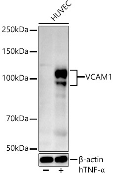
Western blot analysis of lysates from HUVEC cells, using VCAM1 Rabbit pAb (CAB0279) at 1:500 dilution. HUVEC cells were treated with Human Tumor Necrosis Factor-α (hTNF-α) (10 ng/ml) for 16 hours. Secondary antibody: HRP-conjugated Goat anti-Rabbit IgG (H+L) (AS014) at 1:10000 dilution. Lysates/proteins: 25μg per lane. Blocking buffer: 3% nonfat dry milk in TBST. Detection: ECL Basic Kit (AbGn00020). Exposure time: 90s.
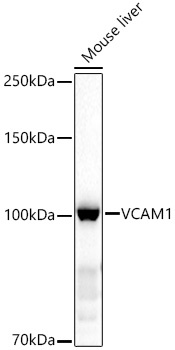
Western blot analysis of lysates from Mouse liver, using VCAM1 Rabbit pAb (CAB0279) at 1:1000 dilution. Secondary antibody: HRP-conjugated Goat anti-Rabbit IgG (H+L) (AS014) at 1:10000 dilution. Lysates/proteins: 25μg per lane. Blocking buffer: 3% nonfat dry milk in TBST. Detection: ECL Basic Kit (AbGn00020). Exposure time: 30s.