The VDAC2 Antibody (CAB18683) is a high-quality antibody developed for reliable detection and analysis of target proteins. VDAC2 is a mitochondrial protein involved in regulating cell metabolism, apoptosis, and cell survival. The antibody, generated in rabbits, is specifically designed for use in Western blot applications and is highly reactive with human samples.By targeting the VDAC2 protein, this antibody allows for the detection and analysis of VDAC2 expression in various cell types, making it ideal for research in mitochondrial biology, cell death pathways, and cancer research.
This antibody is validated for use in WB, IHC-P, IF/ICC, ELISA applications and has demonstrated reactivity against Human, Mouse, Rat samples.
Product Name:
VDAC2 Antibody
SKU:
CAB18683
Size:
20μL, 100μL
Reactivity:
Human, Mouse, Rat
Immunogen:
Synthetic peptide. This information is considered to be commercially sensitive.
Recommended starting concentration is 1 μg/mL. Please optimize the concentration based on your specific assay requirements.
Synonyms:
POR, VDAC2
Positive Sample:
HepG2, HeLa, U-251MG, Mouse testis, Mouse brain, Mouse kidney, Rat brain, Rat kidney
Cellular Localization:
Mitochondrion Outer Membrane.
Calculated MW:
32kDa
Observed MW:
35kDa
This gene encodes a member of the voltage-dependent anion channel pore-forming family of proteins that are considered the main pathway for metabolite diffusion across the mitochondrial outer membrane. The encoded protein is also thought to be involved in the mitochondrial apoptotic pathway via regulation of BCL2-antagonist/killer 1 protein activity. Pseudogenes have been identified on chromosomes 1, 2, 12 and 21, and alternative splicing results in multiple transcript variants.
Purification Method
Affinity purification
Gene ID
7417
RRID
AB_2862419
Buffer Information
Store at -20℃. Avoid freeze / thaw cycles. Buffer: PBS containing 50% glycerol, preserved with proclin300 or sodium azide, pH 7.3.
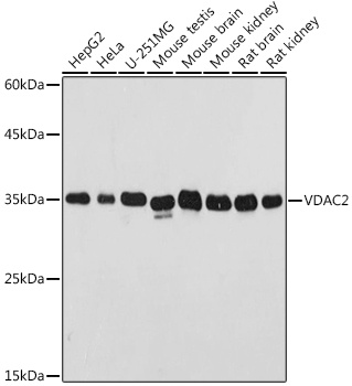
Western blot analysis of various lysates using VDAC2 Rabbit pAb (CAB18683) at 1:500 dilution. Secondary antibody: HRP-conjugated Goat anti-Rabbit IgG (H+L) (CABS014) at 1:10000 dilution. Lysates/proteins: 25μg per lane. Blocking buffer: 3% nonfat dry milk in TBST. Detection: ECL Basic Kit (AbGn00020). Exposure time: 1s.
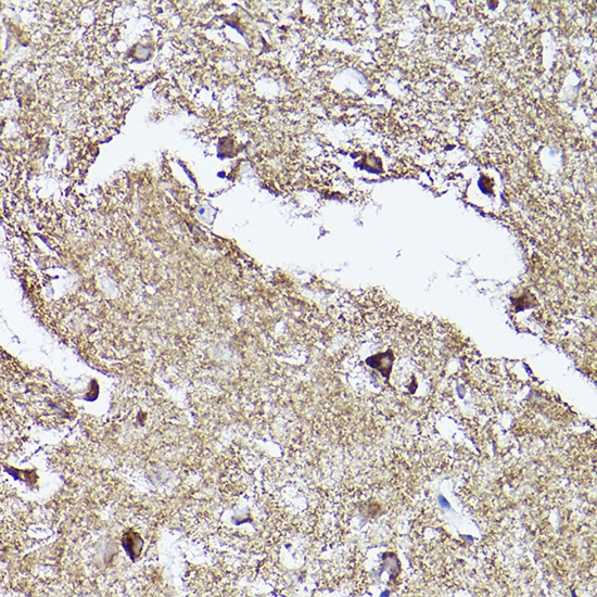
Immunohistochemistry analysis of paraffin-embedded Human brain using VDAC2 Rabbit pAb (CAB18683) at dilution of 1:100 (40x lens). High pressure antigen retrieval performed with 0.01M Citrate buffer (pH 6.0) prior to IHC staining.
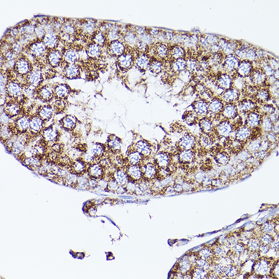
Immunohistochemistry analysis of paraffin-embedded Mouse testis using VDAC2 Rabbit pAb (CAB18683) at dilution of 1:100 (40x lens). High pressure antigen retrieval performed with 0.01M Citrate buffer (pH 6.0) prior to IHC staining.
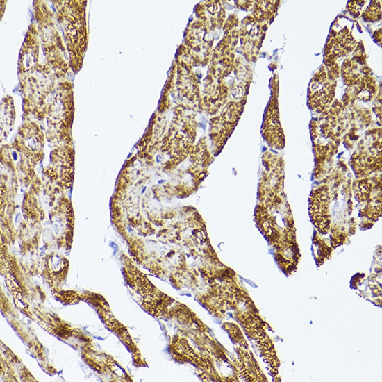
Immunohistochemistry analysis of paraffin-embedded Rat heart using VDAC2 Rabbit pAb (CAB18683) at dilution of 1:100 (40x lens). High pressure antigen retrieval performed with 0.01M Citrate buffer (pH 6.0) prior to IHC staining.
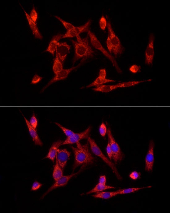
Immunofluorescence analysis of PC-12 cells using VDAC2 Rabbit pAb (CAB18683) at dilution of 1:100 (40x lens). Secondary antibody: Cy3-conjugated Goat anti-Rabbit IgG (H+L) (CABS007) at 1:500 dilution. Blue: DAPI for nuclear staining.


 at 1:10000 dilution. Lysates/proteins: 25ug per lane. Blocking buffer: 3% nonfat dry milk in TBST. Detection: ECL Basic Kit. Exposure time: 60s.")


