The VEGFB Antibody (CAB12689) is a high-quality antibody developed for reliable detection and analysis of target proteins. This antibody, generated in rabbits, is highly specific for detecting VEGFB in human samples and is validated for use in Western blot applications. By binding to the VEGFB protein, this antibody enables the visualization and analysis of VEGFB expression in various cell types, making it useful for investigations in vascular biology and cancer research.VEGFB is a member of the VEGF family known for its role in promoting blood vessel growth and enhancing vascular function.
This antibody is validated for use in WB, IHC-P, ELISA applications and has demonstrated reactivity against Human, Mouse, Rat samples.
Product Name:
VEGFB Antibody
SKU:
CAB12689
Size:
20μL, 100μL
Reactivity:
Human, Mouse, Rat
Conjugate:
Unconjugated
Immunogen:
Synthetic peptide. This information is considered to be commercially sensitive.
Recommended starting concentration is 1 μg/mL. Please optimize the concentration based on your specific assay requirements.
Synonyms:
VRF, VEGFL, VEGFB
Positive Sample:
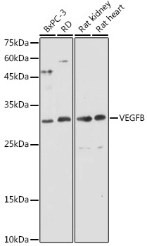
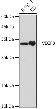
BxPC-3, RD, Rat kidney, Rat heart
Cellular Localization:
Secreted.
Calculated MW:
22kDa
Observed MW:
32kDa
This gene encodes a member of the PDGF (platelet-derived growth factor)/VEGF (vascular endothelial growth factor) family. The VEGF family members regulate the formation of blood vessels and are involved in endothelial cell physiology. This member is a ligand for VEGFR-1 (vascular endothelial growth factor receptor 1) and NRP-1 (neuropilin-1). Studies in mice showed that this gene was co-expressed with nuclear-encoded mitochondrial genes and the encoded protein specifically controlled endothelial uptake of fatty acids. Alternatively spliced transcript variants encoding distinct isoforms have been identified.
Purification Method
Affinity purification
Gene ID
7423
RRID
AB_2759532
Buffer Information
Store at -20℃. Avoid freeze / thaw cycles. Buffer: PBS with 0.01% thimerosal,50% glycerol,pH7.3.
Western blot analysis of various lysates using VEGFB Rabbit pAb (CAB12689) at 1:1000 dilution. Secondary antibody: HRP-conjugated Goat anti-Rabbit IgG (H+L) (CABS014) at 1:10000 dilution. Lysates/proteins: 25μg per lane. Blocking buffer: 3% nonfat dry milk in TBST. Detection: ECL Basic Kit (AbGn00020). Exposure time: 30s.
Western blot analysis of various lysates using VEGFB Rabbit pAb (CAB12689) at 1:1000 dilution. Secondary antibody: HRP-conjugated Goat anti-Rabbit IgG (H+L) (CABS014) at 1:10000 dilution. Lysates/proteins: 25μg per lane. Blocking buffer: 3% nonfat dry milk in TBST. Detection: ECL Basic Kit (AbGn00020). Exposure time: 5s.
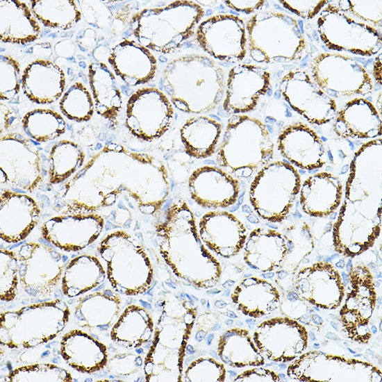
Immunohistochemistry analysis of paraffin-embedded Rat kidney using VEGFB Rabbit pAb (CAB12689) at dilution of 1:100 (40x lens). High pressure antigen retrieval performed with 0.01M Citrate buffer (pH 6.0) prior to IHC staining.
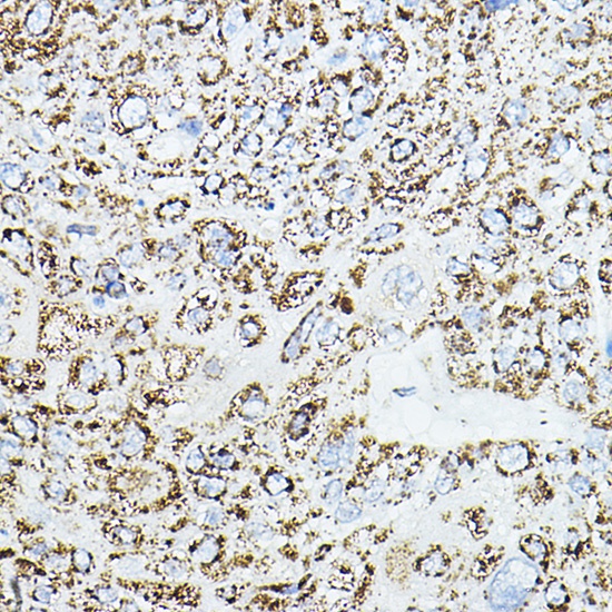
Immunohistochemistry analysis of paraffin-embedded Human esophageal cancer using VEGFB Rabbit pAb (CAB12689) at dilution of 1:100 (40x lens). High pressure antigen retrieval performed with 0.01M Citrate buffer (pH 6.0) prior to IHC staining.


 mAb (HDBS0687)")
 mAb (HDBS1056)")

")