The VHL Antibody (CAB0377) is a high-quality antibody developed for reliable detection and analysis of target proteins. This antibody, produced in rabbits, is highly specific to human VHL protein and has been validated for use in Western blot applications. By binding to the VHL protein, this antibody enables the detection and analysis of VHL in various cell types, making it an ideal choice for studies in cancer research and molecular biology.The VHL protein is known for its role in regulating the stability of hypoxia-inducible factors (HIFs) and plays a crucial role in oxygen sensing and angiogenesis.
This antibody is validated for use in WB, IHC-P, IF/ICC, ELISA applications and has demonstrated reactivity against Human, Mouse, Rat samples.
Product Name:
VHL Antibody
SKU:
CAB0377
Size:
20μL, 100μL
Reactivity:
Human, Mouse, Rat
Conjugate:
Unconjugated
Immunogen:
Synthetic peptide. This information is considered to be commercially sensitive.
This gene encodes a component of a ubiquitination complex. The encoded protein is involved in the ubiquitination and degradation of hypoxia-inducible-factor (HIF), which is a transcription factor that plays a central role in the regulation of gene expression by oxygen. In addition to oxygen-related gene expression, this protein plays a role in many other cellular processes including cilia formation, cytokine signaling, regulation of senescence, and formation of the extracellular matrix. Variants of this gene are associated with von Hippel-Lindau syndrome, pheochromocytoma, erythrocytosis, renal cell carcinoma, and cerebellar hemangioblastoma.
Purification Method
Affinity purification
Gene ID
7428
RRID
AB_2757158
Buffer Information
Store at -20℃. Avoid freeze / thaw cycles. Buffer: PBS containing 50% glycerol, preserved with proclin300 or sodium azide, pH 7.3.
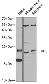
Western blot analysis of various lysates using VHL Rabbit pAb (CAB0377) at 1:1000 dilution. Secondary antibody: HRP-conjugated Goat anti-Rabbit IgG (H+L) (CABS014) at 1:10000 dilution. Lysates/proteins: 25μg per lane. Blocking buffer: 3% nonfat dry milk in TBST. Detection: ECL Basic Kit (AbGn00020). Exposure time: 30s.
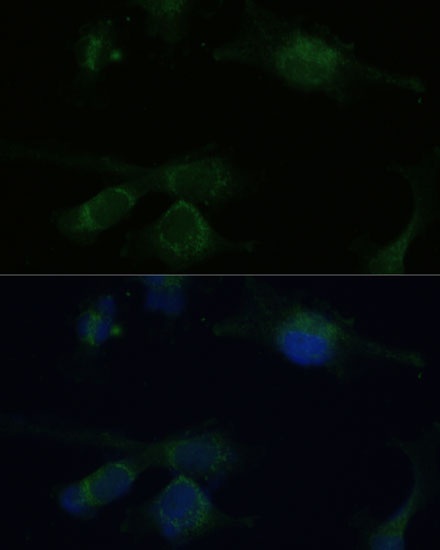
Immunofluorescence analysis of U-251 MG cells using VHL Rabbit pAb (CAB0377) at dilution of 1:100 (40x lens). Secondary antibody: Cy3-conjugated Goat anti-Rabbit IgG (H+L) (CABS007) at 1:500 dilution. Blue: DAPI for nuclear staining.