The VIP Antibody (CAB12531) is a high-quality antibody developed for reliable detection and analysis of target proteins. The protein encoded by this gene belongs to the glucagon family. It stimulates myocardial contractility, causes vasodilation, increases glycogenolysis, lowers arterial blood pressure and relaxes the smooth muscle of trachea, stomach and gall bladder. The protein also acts as an antimicrobial peptide with antibacterial and antifungal activity. Alternative splicing occurs at this locus and two transcript variants encoding distinct isoforms have been identified.
This antibody is validated for use in WB, IHC-P, ELISA applications and has demonstrated reactivity against Human, Mouse, Rat samples.
Product Name:
VIP Antibody
SKU:
CAB12531
Size:
100μL, 20μL
Reactivity:
Human, Mouse, Rat
Conjugate:
Unconjugated
Immunogen:
Recombinant protein (or fragment).This information is considered to be commercially sensitive.
Tested Applications:
WBIHC-PELISA
Recommended Dilution:
WB
1:500 - 1:2000
IHC-P
1:50 - 1:200
ELISA
Recommended starting concentration is 1 μg/mL. Please optimize the concentration based on your specific assay requirements.
Synonyms:
PHM27, VIP
Positive Sample:
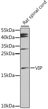
Rat spinal cord
Cellular Localization:
Secreted.
Calculated MW:
19kDa
Observed MW:
19kDa
The protein encoded by this gene belongs to the glucagon family. It stimulates myocardial contractility, causes vasodilation, increases glycogenolysis, lowers arterial blood pressure and relaxes the smooth muscle of trachea, stomach and gall bladder. The protein also acts as an antimicrobial peptide with antibacterial and antifungal activity. Alternative splicing occurs at this locus and two transcript variants encoding distinct isoforms have been identified.
Purification Method
Affinity purification
Gene ID
7432
RRID
AB_2759372
Buffer Information
Store at -20℃. Avoid freeze / thaw cycles. Buffer: PBS containing 50% glycerol, preserved with proclin300 or sodium azide, pH 7.3.
Western blot analysis of lysates from rat spinal cord, using VIP Rabbit pAb (CAB12531) at 1:1000 dilution. Secondary antibody: HRP-conjugated Goat anti-Rabbit IgG (H+L) (AS014) at 1:10000 dilution. Lysates/proteins: 25μg per lane. Blocking buffer: 3% nonfat dry milk in TBST. Detection: ECL Enhanced Kit (AbGn00021). Exposure time: 90s.
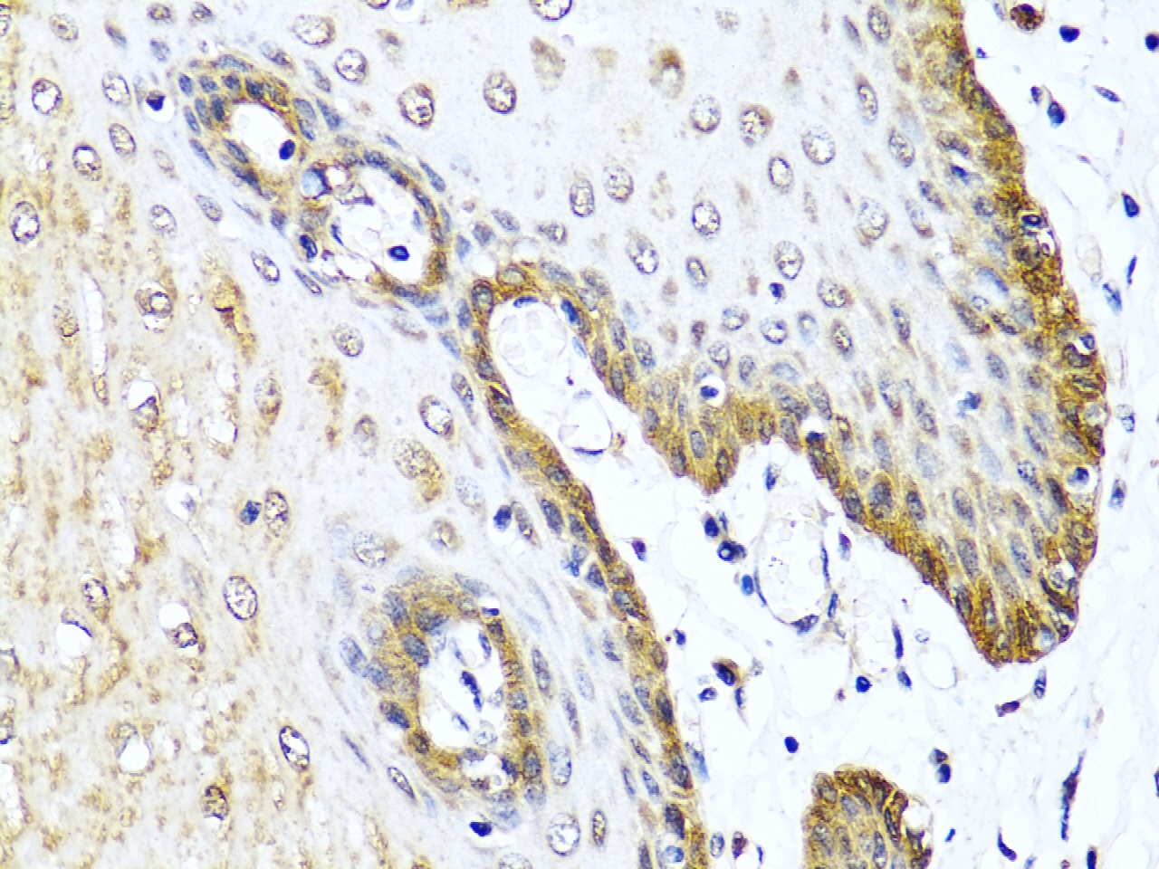
Immunohistochemistry analysis of paraffin-embedded Human esophageal using VIP Rabbit pAb (CAB12531) at dilution of 1:100 (40x lens). Microwave antigen retrieval performed with 0.01M PBS Buffer (pH 7.2) prior to IHC staining.