The WDHD1 Antibody (CAB15396) is a high-quality antibody developed for reliable detection and analysis of target proteins. This antibody is produced in rabbits and has been validated for use in Western blot applications with human samples. It is highly reactive and specific to WDHD1, allowing for accurate detection and analysis in various cellular contexts.WDHD1 is a protein involved in various cellular processes, including DNA repair, transcriptional regulation, and chromatin remodeling. Research has shown that dysregulation of WDHD1 can lead to abnormalities in cell growth and development, making it an important target for investigation in cancer biology and genetic disorders.
This antibody is validated for use in WB, IHC-P, IF/ICC, ELISA applications and has demonstrated reactivity against Human, Rat samples.
Product Name:
WDHD1 Antibody
SKU:
CAB15396
Size:
20μL, 100μL
Reactivity:
Human, Rat
Conjugate:
Unconjugated
Immunogen:
Recombinant protein (or fragment).This information is considered to be commercially sensitive.
Recommended starting concentration is 1 μg/mL. Please optimize the concentration based on your specific assay requirements.
Synonyms:
AND1, CTF4, AND-1, CHTF4, WDHD1
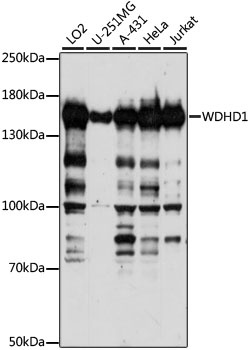
Positive Sample:
LO2, U-251MG, A-431, HeLa, Jurkat
Cellular Localization:
Nucleus, Nucleoplasm.
Calculated MW:
126kDa
Observed MW:
135kDa
The protein encoded by this gene contains multiple N-terminal WD40 domains and a C-terminal high mobility group (HMG) box. WD40 domains are found in a variety of eukaryotic proteins and may function as adaptor/regulatory modules in signal transduction, pre-mRNA processing and cytoskeleton assembly. HMG boxes are found in many eukaryotic proteins involved in chromatin assembly, transcription and replication. Alternative splicing results in two transcript variants encoding different isoforms.
Purification Method
Affinity purification
Gene ID
11169
RRID
AB_2762303
Buffer Information
Store at -20℃. Avoid freeze / thaw cycles. Buffer: PBS with 0.01% thimerosal,50% glycerol,pH7.3.
Western blot analysis of various lysates using WDHD1 Rabbit pAb (CAB15396) at 1:1000 dilution. Secondary antibody: HRP-conjugated Goat anti-Rabbit IgG (H+L) (CABS014) at 1:10000 dilution. Lysates/proteins: 25μg per lane. Blocking buffer: 3% nonfat dry milk in TBST. Detection: ECL Basic Kit (AbGn00020). Exposure time: 10s.
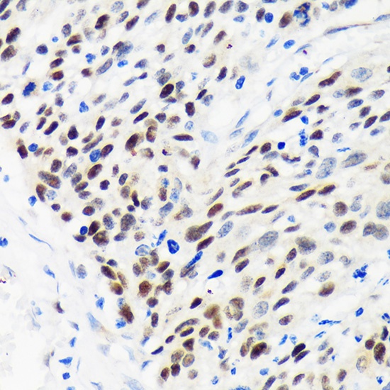
Immunohistochemistry analysis of paraffin-embedded Human esophageal cancer using WDHD1 Rabbit pAb (CAB15396) at dilution of 1:100 (40x lens). Microwave antigen retrieval performed with 0.01M PBS Buffer (pH 7.2) prior to IHC staining.