The WDR5 Monoclonal Antibody (CAB3259) is a high-quality antibody developed for reliable detection and analysis of target proteins. This antibody, raised in rabbits, is highly specific to human samples and has been validated for use in Western blot and immunohistochemistry applications.WDR5 is a crucial component of the MLL complex, which plays a vital role in the maintenance of gene expression patterns and stem cell renewal. Dysregulation of WDR5 has been implicated in various diseases, including cancer and neurological disorders. By targeting WDR5 with this monoclonal antibody, researchers can better understand the mechanisms underlying these diseases and explore potential therapeutic interventions.
This antibody is validated for use in WB, ELISA applications and has demonstrated reactivity against Human, Rat samples.
Product Name:
WDR5 Monoclonal Antibody
SKU:
CAB3259
Size:
20μL, 100μL
Reactivity:
Human, Rat
Clone Number:
ARC0769
Conjugate:
Unconjugated
Immunogen:
Synthetic peptide. This information is considered to be commercially sensitive.
Recommended starting concentration is 1 μg/mL. Please optimize the concentration based on your specific assay requirements.
Synonyms:
BIG3, SWD3, BIG-3, CFAP89, WDR5
Positive Sample:
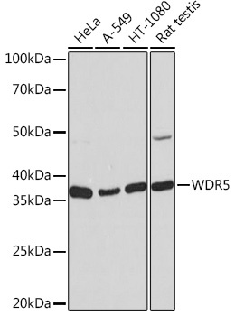
HeLa, A549, HT-1080, Rat testis
Cellular Localization:
Nucleus.
Calculated MW:
37kDa
Observed MW:
37kDa
This gene encodes a member of the WD repeat protein family. WD repeats are minimally conserved regions of approximately 40 amino acids typically bracketed by gly-his and trp-asp (GH-WD), which may facilitate formation of heterotrimeric or multiprotein complexes. Members of this family are involved in a variety of cellular processes, including cell cycle progression, signal transduction, apoptosis, and gene regulation. This protein contains 7 WD repeats. Alternatively spliced transcript variants encoding the same protein have been identified.
Purification Method
Affinity purification
Gene ID
11091
RRID
AB_2863033
Buffer Information
Store at -20℃. Avoid freeze / thaw cycles. Buffer: PBS containing 50% glycerol and 0.05% BSA, preserved with proclin300 or sodium azide, pH 7.3.
Western blot analysis of various lysates using WDR5 Rabbit mAb (CAB3259) at 1:1000 dilution. Secondary antibody: HRP-conjugated Goat anti-Rabbit IgG (H+L) (CABS014) at 1:10000 dilution. Lysates/proteins: 25μg per lane. Blocking buffer: 3% nonfat dry milk in TBST. Detection: ECL Basic Kit (AbGn00020). Exposure time: 10s.