The WIPF1 Antibody (CAB17003) is a high-quality antibody developed for reliable detection and analysis of target proteins. Raised in rabbits, this antibody is highly specific and sensitive to human samples, making it ideal for Western blot applications. By binding to the WIPF1 protein, this antibody allows for the detection and analysis of WIPF1 in various cell types, essential for studies in cell biology, cancer research, and immunology.
This antibody is validated for use in WB, IHC-P, ELISA applications and has demonstrated reactivity against Human, Mouse, Rat samples.
Product Name:
WIPF1 Antibody
SKU:
CAB17003
Size:
20μL, 100μL
Reactivity:
Human, Mouse, Rat
Immunogen:
Synthetic peptide. This information is considered to be commercially sensitive.
Recommended starting concentration is 1 μg/mL. Please optimize the concentration based on your specific assay requirements.
Synonyms:
WIP, WAS2, PRPL-2, WASPIP, WIPF1
Positive Sample:
RAW264.7, Mouse spleen
Cellular Localization:
Actin Cytoskeleton, Cytoplasmic Vesicle, Cytosol.
Calculated MW:
51kDa
Observed MW:
51kDa
This gene encodes a protein that plays an important role in the organization of the actin cytoskeleton. The encoded protein binds to a region of Wiskott-Aldrich syndrome protein that is frequently mutated in Wiskott-Aldrich syndrome, an X-linked recessive disorder. Impairment of the interaction between these two proteins may contribute to the disease. Two transcript variants encoding the same protein have been identified for this gene.
Purification Method
Affinity purification
Gene ID
7456
RRID
AB_2772897
Buffer Information
Store at -20℃. Avoid freeze / thaw cycles. Buffer: PBS with 0.01% thimerosal,50% glycerol,pH7.3.
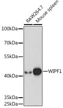
Western blot analysis of various lysates using WIPF1 Rabbit pAb (CAB17003) at 1:1000 dilution. Secondary antibody: HRP-conjugated Goat anti-Rabbit IgG (H+L) (CABS014) at 1:10000 dilution. Lysates/proteins: 25μg per lane. Blocking buffer: 3% nonfat dry milk in TBST. Detection: ECL Basic Kit (AbGn00020). Exposure time: 10s.
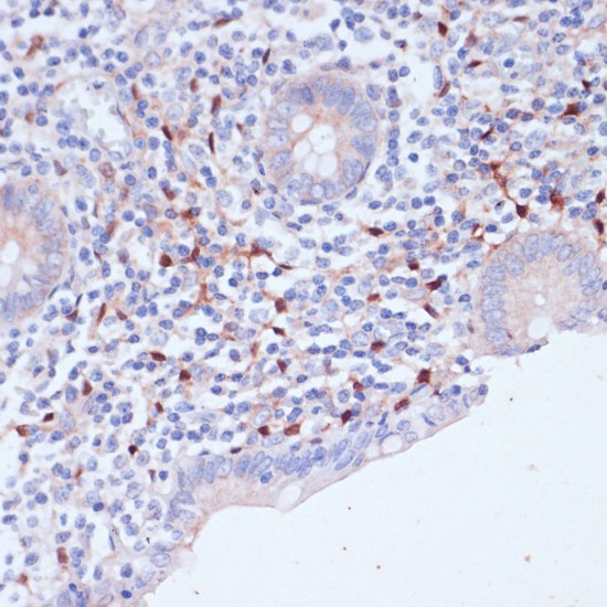
Immunohistochemistry analysis of paraffin-embedded Human appendix using WIPF1 Rabbit pAb (CAB17003) at dilution of 1:100 (40x lens). Microwave antigen retrieval performed with 0.01M PBS Buffer (pH 7.2) prior to IHC staining.
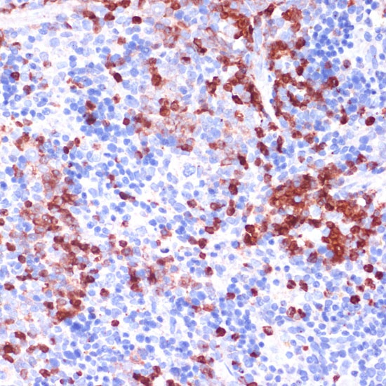
Immunohistochemistry analysis of paraffin-embedded Mouse spleen using WIPF1 Rabbit pAb (CAB17003) at dilution of 1:100 (40x lens). Microwave antigen retrieval performed with 0.01M PBS Buffer (pH 7.2) prior to IHC staining.