The WNT1 Antibody (CAB2475) is a high-quality antibody developed for reliable detection and analysis of target proteins. This antibody, raised in rabbits, is highly specific for WNT1 in human samples and has been validated for use in Western blot applications. By binding to the WNT1 protein, this antibody allows for accurate detection and analysis in a variety of cell types, making it ideal for studies in developmental biology and cancer research.WNT1 is a key player in the Wnt signaling pathway, which is crucial for embryonic development, tissue regeneration, and stem cell maintenance.
This antibody is validated for use in WB, IHC-P, IF/ICC, ELISA applications and has demonstrated reactivity against Human, Mouse, Rat samples.
Product Name:
WNT1 Antibody
SKU:
CAB2475
Size:
20μL, 100μL
Reactivity:
Human, Mouse, Rat
Conjugate:
Unconjugated
Immunogen:
Recombinant protein (or fragment).This information is considered to be commercially sensitive.
The WNT gene family consists of structurally related genes which encode secreted signaling proteins. These proteins have been implicated in oncogenesis and in several developmental processes, including regulation of cell fate and patterning during embryogenesis. This gene is a member of the WNT gene family. It is very conserved in evolution, and the protein encoded by this gene is known to be 98% identical to the mouse Wnt1 protein at the amino acid level. The studies in mouse indicate that the Wnt1 protein functions in the induction of the mesencephalon and cerebellum. This gene was originally considered as a candidate gene for Joubert syndrome, an autosomal recessive disorder with cerebellar hypoplasia as a leading feature. However, further studies suggested that the gene mutations might not have a significant role in Joubert syndrome. This gene is clustered with another family member, WNT10B, in the chromosome 12q13 region.
Purification Method
Affinity purification
Gene ID
7471
RRID
AB_2764374
Buffer Information
Store at -20℃. Avoid freeze / thaw cycles. Buffer: PBS containing 50% glycerol, preserved with proclin300 or sodium azide, pH 7.3.
Western blot analysis of various lysates using WNT1 Rabbit pAb (CAB2475) at 1:8000 dilution. Secondary antibody: HRP-conjugated Goat anti-Rabbit IgG (H+L) (CABS014) at 1:10000 dilution. Lysates / proteins: 25 μg per lane. Blocking buffer: 3 % nonfat dry milk in TBST. Detection: ECL Basic Kit (AbGn00020). Exposure time: 60s.
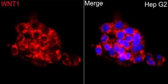
Immunofluorescence analysis of HepG2 cells using WNT1 Rabbit pAb (CAB2475) at dilution of 1:200 (40x lens). Secondary antibody: Cy3-conjugated Goat anti-Rabbit IgG (H+L) (CABS007) at 1:500 dilution. Blue: DAPI for nuclear staining.
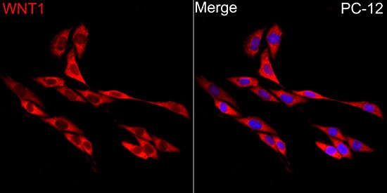
Immunofluorescence analysis of PC-12 cells using WNT1 Rabbit pAb (CAB2475) at dilution of 1:200 (40x lens). Secondary antibody: Cy3-conjugated Goat anti-Rabbit IgG (H+L) (CABS007) at 1:500 dilution. Blue: DAPI for nuclear staining.


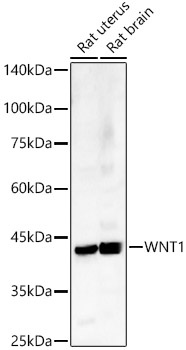


 at 1:10000 dilution. Lysates/proteins: 25ug per lane. Blocking buffer: 3% nonfat dry milk in TBST. Detection: ECL Basic Kit. Exposure time: 10s.")


