The WTAP Antibody (CAB14695) is a high-quality antibody developed for reliable detection and analysis of target proteins. This antibody, produced in rabbits, exhibits high reactivity with human samples and has been validated for use in Western blot applications. By specifically binding to the WTAP protein, this antibody enables accurate detection and analysis in a variety of cell types, making it an ideal choice for studies in molecular biology and cancer research.
This antibody is validated for use in WB, IF/ICC, ELISA applications and has demonstrated reactivity against Human, Mouse, Rat samples.
Product Name:
WTAP Antibody
SKU:
CAB14695
Size:
20μL, 100μL
Reactivity:
Human, Mouse, Rat
Conjugate:
Unconjugated
Immunogen:
Recombinant protein (or fragment).This information is considered to be commercially sensitive.
Recommended starting concentration is 1 μg/mL. Please optimize the concentration based on your specific assay requirements.
Synonyms:
Mum2, WTAP
Positive Sample:
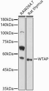
RAW264.7, Rat thymus
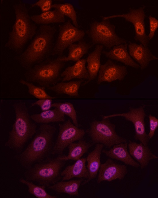
Cellular Localization:
Nucleus, Nucleus Speckle, Nucleoplasm.
Calculated MW:
44kDa
Observed MW:
55kDa
The Wilms tumor suppressor gene WT1 appears to play a role in both transcriptional and posttranscriptional regulation of certain cellular genes. This gene encodes a WT1-associating protein, which is a ubiquitously expressed nuclear protein. Like WT1 protein, this protein is localized throughout the nucleoplasm as well as in speckles and partially colocalizes with splicing factors. Alternative splicing of this gene results in several transcript variants encoding three different isoforms.
Purification Method
Affinity purification
Gene ID
9589
RRID
AB_2761570
Buffer Information
Store at -20℃. Avoid freeze / thaw cycles. Buffer: PBS containing 50% glycerol, preserved with proclin300 or sodium azide, pH 7.3.
Western blot analysis of various lysates using WTAP Rabbit pAb (CAB14695) at 1:1000 dilution. Secondary antibody: HRP-conjugated Goat anti-Rabbit IgG (H+L) (CABS014) at 1:10000 dilution. Lysates/proteins: 25μg per lane. Blocking buffer: 3% nonfat dry milk in TBST. Detection: ECL Basic Kit (AbGn00020). Exposure time: 30s.
Immunofluorescence analysis of HeLa cells using WTAP Rabbit pAb (CAB14695) at dilution of 1:50 (40x lens). Secondary antibody: Cy3-conjugated Goat anti-Rabbit IgG (H+L) (CABS007) at 1:500 dilution. Blue: DAPI for nuclear staining.