The XK Antibody (CAB15109) is a high-quality antibody developed for reliable detection and analysis of target proteins. Raised in rabbits, this antibody is highly specific and reactive with human samples, providing reliable results in Western blot applications.XK is essential for the transport of various ions and molecules across the red blood cell membrane, making it crucial for normal blood cell function. Dysregulation of XK has been linked to various blood disorders, including Kell antigen deficiency and McLeod syndrome.
This antibody is validated for use in WB, IF/ICC, ELISA applications and has demonstrated reactivity against Human, Mouse, Rat samples.
Product Name:
XK Antibody
SKU:
CAB15109
Size:
20μL, 100μL
Reactivity:
Human, Mouse, Rat
Conjugate:
Unconjugated
Immunogen:
Recombinant protein (or fragment).This information is considered to be commercially sensitive.
Recommended starting concentration is 1 μg/mL. Please optimize the concentration based on your specific assay requirements.
Synonyms:
KX, NA, NAC, X1k, XKR1, XK
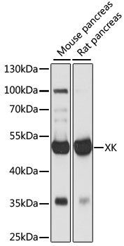
Positive Sample:
mouse pancreas, rat pancreas
Cellular Localization:
Membrane, Multi-Pass Membrane Protein.
Calculated MW:
51kDa
Observed MW:
50kDa
This locus controls the synthesis of the Kell blood group 'precursor substance' (Kx). Mutations in this gene have been associated with McLeod syndrome, an X-linked, recessive disorder characterized by abnormalities in the neuromuscular and hematopoietic systems. The encoded protein has structural characteristics of prokaryotic and eukaryotic membrane transport proteins.
Purification Method
Affinity purification
Gene ID
7504
RRID
AB_2761993
Buffer Information
Store at -20℃. Avoid freeze / thaw cycles. Buffer: PBS with 0.01% thimerosal,50% glycerol,pH7.3.
Western blot analysis of various lysates using XK Rabbit pAb (CAB15109) at 1:1000 dilution. Secondary antibody: HRP-conjugated Goat anti-Rabbit IgG (H+L) (CABS014) at 1:10000 dilution. Lysates/proteins: 25μg per lane. Blocking buffer: 3% nonfat dry milk in TBST. Detection: ECL Basic Kit (AbGn00020). Exposure time: 1s.
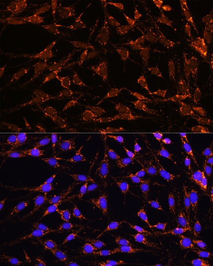
Immunofluorescence analysis of C6 cells using XK Rabbit pAb (CAB15109) at dilution of 1:100. Secondary antibody: Cy3-conjugated Goat anti-Rabbit IgG (H+L) (CABS007) at 1:500 dilution. Blue: DAPI for nuclear staining.
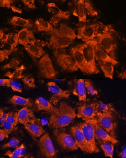
Immunofluorescence analysis of U-2 OS cells using XK Rabbit pAb (CAB15109) at dilution of 1:100. Secondary antibody: Cy3-conjugated Goat anti-Rabbit IgG (H+L) (CABS007) at 1:500 dilution. Blue: DAPI for nuclear staining.