The YAP1 Monoclonal Antibody (CAB19134) is a high-quality antibody developed for reliable detection and analysis of target proteins. This antibody, raised in rabbits, exhibits high reactivity with human samples and has been validated for use in various applications, including Western blot and immunohistochemistry.YAP1 is a critical regulator of cellular growth and survival, and dysregulation of its activity has been implicated in the development and progression of various cancers, making it a promising target for cancer research.
This antibody is validated for use in WB, IHC-P, IF/ICC, IP, ELISA applications and has demonstrated reactivity against Human, Mouse, Rat samples.
Product Name:
YAP1 Monoclonal Antibody
SKU:
CAB19134
Size:
20μL, 100μL
Reactivity:
Human, Mouse, Rat
Clone Number:
ARC53477
Conjugate:
Unconjugated
Immunogen:
Recombinant protein (or fragment).This information is considered to be commercially sensitive.
0.5μg-4μg antibody for 200μg-400μg extracts of whole cells
ELISA
Recommended starting concentration is 1 μg/mL. Please optimize the concentration based on your specific assay requirements.
Synonyms:
YAP, YKI, COB1, YAP2, YAP-1, YAP65, P1
Positive Sample:
HeLa, Mouse heart, A549
Cellular Localization:
Cytoplasm, Nucleus.
Calculated MW:
36kDa-54kDa
Observed MW:
70kDa
This gene encodes a downstream nuclear effector of the Hippo signaling pathway which is involved in development, growth, repair, and homeostasis. This gene is known to play a role in the development and progression of multiple cancers as a transcriptional regulator of this signaling pathway and may function as a potential target for cancer treatment. Alternative splicing results in multiple transcript variants encoding different isoforms.
Purification Method
Affinity purification
Gene ID
10413
RRID
AB_2862627
Buffer Information
Store at -20℃. Avoid freeze / thaw cycles. Buffer: PBS containing 50% glycerol and 0.05% BSA, preserved with proclin300 or sodium azide, pH 7.3.
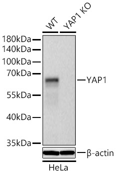
Western blot analysis of lysates from wild type (WT) and YAP1 knockout (KO) HeLa cells using [KO Validated] YAP1 Rabbit mAb (CAB19134) at 1:10000 dilution incubated overnight at 4℃. Secondary antibody: HRP-conjugated Goat anti-Rabbit IgG (H+L) (CABS014) at 1:10000 dilution. Lysates/proteins: 25 μg per lane. Blocking buffer: 3% nonfat dry milk in TBST. Detection: ECL Basic Kit (AbGn00020). Exposure time: 10s.
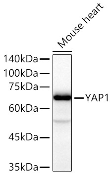
Western blot analysis of lysates from Mouse heart using [KO Validated] YAP1 Rabbit mAb (CAB19134) at 1:20000 dilution incubated overnight at 4℃. Secondary antibody: HRP-conjugated Goat anti-Rabbit IgG (H+L) (CABS014) at 1:10000 dilution. Lysates/proteins: 25 μg per lane. Blocking buffer: 3% nonfat dry milk in TBST. Detection: ECL Basic Kit (AbGn00020). Exposure time: 20 s.
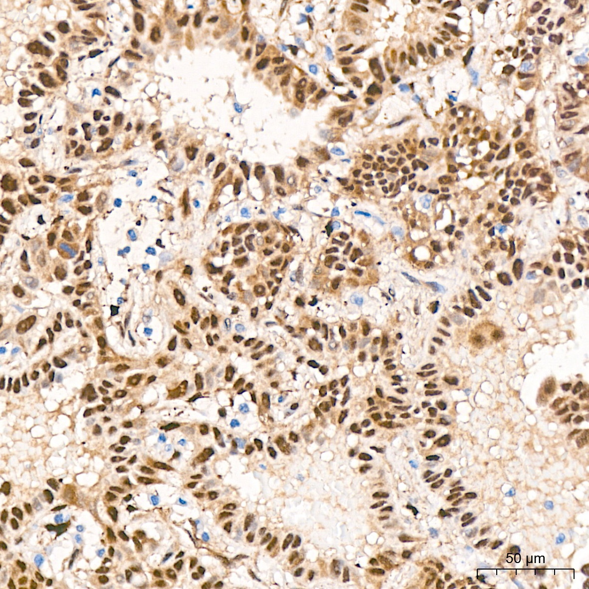
Immunohistochemistry analysis of paraffin-embedded Human lung adenocarcinoma tissue using [KO Validated] YAP1 Rabbit mAb (CAB19134) at a dilution of 1:400 (40x lens). High pressure antigen retrieval performed with 0.01M Citrate buffer (pH 6.0) prior to IHC staining.
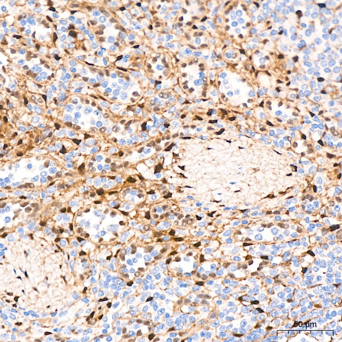
Immunohistochemistry analysis of paraffin-embedded Human spleen tissue using [KO Validated] YAP1 Rabbit mAb (CAB19134) at a dilution of 1:400 (40x lens). High pressure antigen retrieval performed with 0.01M Citrate buffer (pH 6.0) prior to IHC staining.
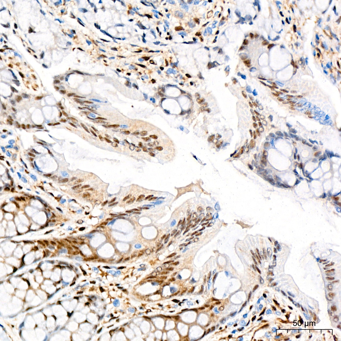
Immunohistochemistry analysis of paraffin-embedded Rat colon tissue using [KO Validated] YAP1 Rabbit mAb (CAB19134) at a dilution of 1:400 (40x lens). High pressure antigen retrieval performed with 0.01M Citrate buffer (pH 6.0) prior to IHC staining.
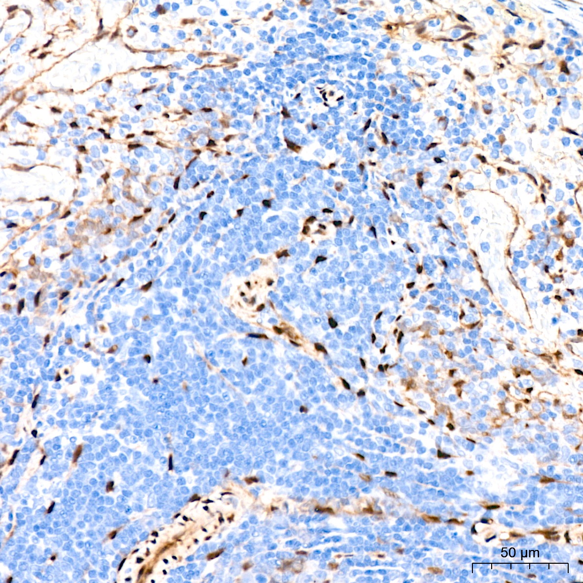
Immunohistochemistry analysis of paraffin-embedded Rat spleen tissue using [KO Validated] YAP1 Rabbit mAb (CAB19134) at a dilution of 1:400 (40x lens). High pressure antigen retrieval performed with 0.01M Citrate buffer (pH 6.0) prior to IHC staining.
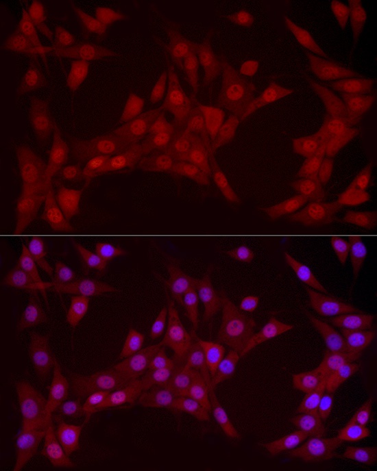
Immunofluorescence analysis of NIH/3T3 cells using [KO Validated] YAP1 Rabbit mAb (CAB19134) at dilution of 1:100 (40x lens). Secondary antibody: Cy3-conjugated Goat anti-Rabbit IgG (H+L) (CABS007) at 1:500 dilution. Blue: DAPI for nuclear staining.
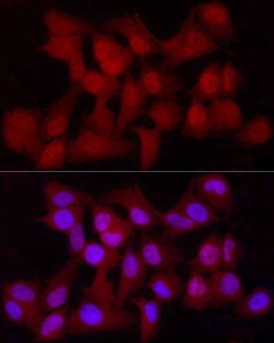
Immunofluorescence analysis of HeLa cells using [KO Validated] YAP1 Rabbit mAb (CAB19134) at dilution of 1:100 (40x lens). Secondary antibody: Cy3-conjugated Goat anti-Rabbit IgG (H+L) (CABS007) at 1:500 dilution. Blue: DAPI for nuclear staining.
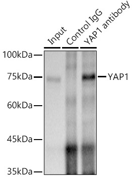
Immunoprecipitation analysis of 300 μg extracts of A-549 cells using 3 μg [KO Validated] YAP1 Rabbit mAb (CAB19134). Western blot was performed from the immunoprecipitate using [KO Validated] YAP1 Rabbit mAb(CAB19134) at a dilution of 1:500.


")


