The YTHDF1 Polyclonal Antibody (CAB13260) is a high-quality antibody developed for reliable detection and analysis of target proteins. This antibody, produced in rabbits, exhibits high specificity and sensitivity for detecting YTHDF1 in human samples, making it suitable for various applications such as Western blot and immunohistochemistry.YTHDF1 is a member of the YTH domain-containing family of proteins, which play important roles in mRNA decay and translation regulation. As a reader protein for N6-methyladenosine (m6A) modifications on RNA molecules, YTHDF1 is involved in various biological processes, including cell differentiation, development, and cancer progression.
This antibody is validated for use in WB, IF/ICC, IP, ELISA applications and has demonstrated reactivity against Human, Mouse, Rat samples.
Product Name:
YTHDF1 Polyclonal Antibody
SKU:
CAB13260
Size:
20μL, 100μL
Reactivity:
Human, Mouse, Rat
Conjugate:
Unconjugated
Immunogen:
Recombinant protein (or fragment).This information is considered to be commercially sensitive.
Recommended starting concentration is 1 μg/mL. Please optimize the concentration based on your specific assay requirements.
Synonyms:
DF1, C20orf21, YTHDF1
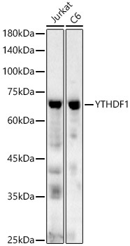
Positive Sample:
Jurkat, C6
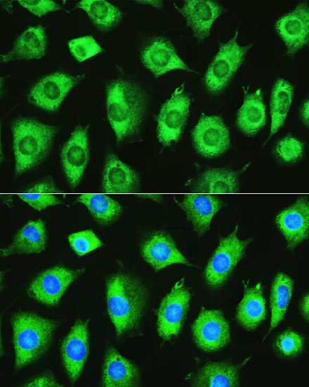
Cellular Localization:
Cytoplasm.
Calculated MW:
61kDa
Observed MW:
70kDa
Enables N6-methyladenosine-containing RNA binding activity and ribosome binding activity. Involved in mRNA destabilization; positive regulation of translational initiation; and stress granule assembly. Located in P-body and cytoplasmic stress granule.
Purification Method
Affinity purification
Gene ID
54915
RRID
AB_2760113
Buffer Information
Store at -20℃. Avoid freeze / thaw cycles. Buffer: Buffer: PBS containing 50% glycerol, preserved with proclin300 or sodium azide, pH 7.3.
Western blot analysis of various lysates, using YTHDF1 Rabbit pAb (CAB13260) at 1:500 dilution. Secondary antibody: HRP-conjugated Goat anti-Rabbit IgG (H+L) (CABS014) at 1:10000 dilution. Lysates/proteins: 25μg per lane. Blocking buffer: 3% nonfat dry milk in TBST. Detection: ECL Basic Kit (AbGn00020). Exposure time: 60s.
Immunofluorescence analysis of L929 cells using YTHDF1 Rabbit pAb (CAB13260) at dilution of 1:100. Secondary antibody: Cy3-conjugated Goat anti-Rabbit IgG (H+L) (CABS007) at 1:500 dilution. Blue: DAPI for nuclear staining.