The ACAA1 Antibody (CAB7422) is a high-quality antibody developed for reliable detection and analysis of target proteins. This antibody, produced in rabbits, exhibits high reactivity with human samples and has been validated for use in Western blot applications. By binding specifically to the ACAA1 protein, researchers can accurately detect and analyze ACAA1 expression in various cell types, making it an essential reagent for studies in metabolism and lipid biology.ACAA1 is a key enzyme in the beta-oxidation pathway, playing a crucial role in breaking down fatty acids for energy production. Dysregulation of ACAA1 has been linked to metabolic disorders, including obesity and fatty liver disease.
This antibody is validated for use in WB, ELISA applications and has demonstrated reactivity against Human, Rat samples.
Product Name:
ACAA1 Antibody
SKU:
CAB7422
Size:
20μL, 100μL
Reactivity:
Human, Rat
Conjugate:
Unconjugated
Immunogen:
Recombinant protein (or fragment).This information is considered to be commercially sensitive.
Recommended starting concentration is 1 μg/mL. Please optimize the concentration based on your specific assay requirements.
Synonyms:
ACAA, THIO, PTHIO, Lnc-Myd88, ACAA1
Positive Sample:
HL-60, MCF7, A-549, Jurkat
Cellular Localization:
Peroxisome.
Calculated MW:
44kDa
Observed MW:
44kDa
This gene encodes an enzyme operative in the beta-oxidation system of the peroxisomes. Deficiency of this enzyme leads to pseudo-Zellweger syndrome. Alternative splicing results in multiple transcript variants.
Purification Method
Affinity purification
Gene ID
30
RRID
AB_2767950
Buffer Information
Store at -20℃. Avoid freeze / thaw cycles. Buffer: PBS containing 50% glycerol, preserved with proclin300 or sodium azide, pH 7.3.
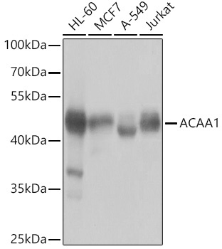
Western blot analysis of various lysates using ACAA1 Rabbit pAb (CAB7422) at 1:1000 dilution. Secondary antibody: HRP-conjugated Goat anti-Rabbit IgG (H+L) (CABS014) at 1:10000 dilution. Lysates/proteins: 25μg per lane. Blocking buffer: 3% nonfat dry milk in TBST. Detection: ECL Basic Kit (AbGn00020). Exposure time: 1s.
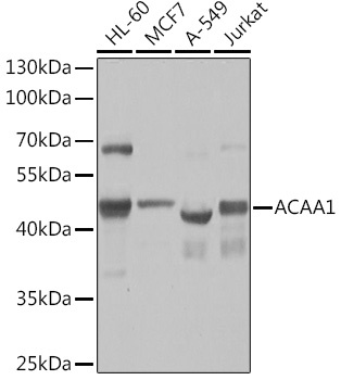
Western blot analysis of various lysates using ACAA1 Rabbit pAb (CAB7422) at 1:1000 dilution. Secondary antibody: HRP-conjugated Goat anti-Rabbit IgG (H+L) (CABS014) at 1:10000 dilution. Lysates/proteins: 25μg per lane. Blocking buffer: 3% nonfat dry milk in TBST. Detection: ECL Basic Kit (AbGn00020). Exposure time: 1s.