The ACAA2 Antibody (CAB15778) is a high-quality antibody developed for reliable detection and analysis of target proteins. This antibody, generated in rabbits, is highly specific and reacts well with human samples, making it ideal for Western blot experiments. By binding to the ACAA2 protein, this antibody enables the detection and analysis of ACAA2 expression in various cell types, allowing for detailed investigation into its role in lipid metabolism and related diseases.
This antibody is validated for use in WB, IHC-P, IF/ICC, ELISA applications and has demonstrated reactivity against Human, Mouse, Rat samples.
Product Name:
ACAA2 Antibody
SKU:
CAB15778
Size:
20μL, 100μL
Reactivity:
Human, Mouse, Rat
Conjugate:
Unconjugated
Immunogen:
Recombinant protein (or fragment).This information is considered to be commercially sensitive.
Recommended starting concentration is 1 μg/mL. Please optimize the concentration based on your specific assay requirements.
Synonyms:
DSAEC, ACAA2
Positive Sample:
Hep G2, HeLa, Mouse liver
Cellular Localization:
Mitochondrion.
Calculated MW:
42kDa
Observed MW:
42kDa
The encoded protein catalyzes the last step of the mitochondrial fatty acid beta-oxidation spiral. Unlike most mitochondrial matrix proteins, it contains a non-cleavable amino-terminal targeting signal.
Purification Method
Affinity purification
Gene ID
10449
RRID
AB_2763197
Buffer Information
Store at -20℃. Avoid freeze / thaw cycles. Buffer: PBS containing 50% glycerol, preserved with proclin300 or sodium azide, pH 7.3.
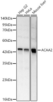
Western blot analysis of various lysates using ACAA2 Rabbit pAb (CAB15778) at 1:1000 dilution. Secondary antibody: HRP-conjugated Goat anti-Rabbit IgG (H+L) (CABS014) at 1:10000 dilution. Lysates / proteins: 25 μg per lane. Blocking buffer: 3 % nonfat dry milk in TBST. Detection: ECL Basic Kit (AbGn00020). Exposure time: 0.8s.
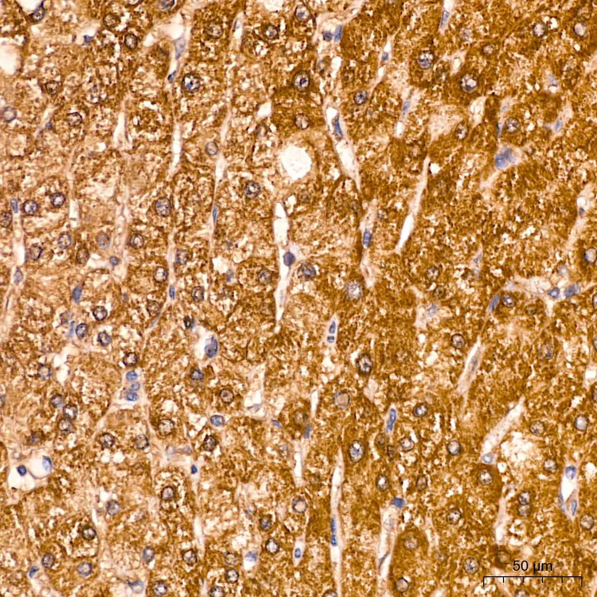
Immunohistochemistry analysis of paraffin-embedded Human liver tissue using ACAA2 Rabbit pAb (CAB15778) at a dilution of 1:100 (40x lens). High pressure antigen retrieval was performed with 0.01 M citrate buffer (pH 6.0) prior to IHC staining.
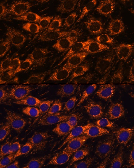
Immunofluorescence analysis of C6 cells using ACAA2 Rabbit pAb (CAB15778) at dilution of 1:100. Secondary antibody: Cy3-conjugated Goat anti-Rabbit IgG (H+L) (CABS007) at 1:500 dilution. Blue: DAPI for nuclear staining.
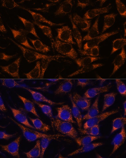
Immunofluorescence analysis of L929 cells using ACAA2 Rabbit pAb (CAB15778) at dilution of 1:100. Secondary antibody: Cy3-conjugated Goat anti-Rabbit IgG (H+L) (CABS007) at 1:500 dilution. Blue: DAPI for nuclear staining.