The ACAT2 Antibody (CAB7866) is a high-quality antibody developed for reliable detection and analysis of target proteins. This antibody is produced through immunization of rabbits and has high specificity for human samples, making it ideal for studies focusing on cholesterol homeostasis and lipid metabolism. ACAT2 is primarily found in the liver and intestine, where it plays a crucial role in the synthesis of cholesterol esters. Dysregulation of ACAT2 activity has been linked to various metabolic disorders, including atherosclerosis and non-alcoholic fatty liver disease.
This antibody is validated for use in WB, IHC-P, IF/ICC, ELISA applications and has demonstrated reactivity against Human, Mouse, Rat samples.
Product Name:
ACAT2 Antibody
SKU:
CAB7866
Size:
20μL, 100μL
Reactivity:
Human, Mouse, Rat
Conjugate:
Unconjugated
Immunogen:
Synthetic peptide. This information is considered to be commercially sensitive.
Recommended starting concentration is 1 μg/mL. Please optimize the concentration based on your specific assay requirements.
Synonyms:
ACAT2
Positive Sample:
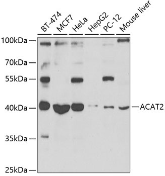
BT-474, MCF7, HeLa, HepG2, PC-12, Mouse liver
Cellular Localization:
Cytoplasm.
Calculated MW:
41kDa
Observed MW:
41kDa
The product of this gene is an enzyme involved in lipid metabolism, and it encodes cytosolic acetoacetyl-CoA thiolase. This gene shows complementary overlapping with the 3-prime region of the TCP1 gene in both mouse and human. These genes are encoded on opposite strands of DNA, as well as in opposite transcriptional orientation. Alternatively spliced transcript variants encoding different isoforms have been found for this gene.
Purification Method
Affinity purification
Gene ID
39
RRID
AB_2768199
Buffer Information
Store at -20℃. Avoid freeze / thaw cycles. Buffer: PBS with 0.09% Sodium azide,50% glycerol,pH7.3.
Western blot analysis of various lysates using ACAT2 Rabbit pAb (CAB7866) at 1:1000 dilution. Secondary antibody: HRP-conjugated Goat anti-Rabbit IgG (H+L) (CABS014) at 1:10000 dilution. Lysates/proteins: 25μg per lane. Blocking buffer: 3% nonfat dry milk in TBST. Detection: ECL Basic Kit (AbGn00020). Exposure time: 90s.
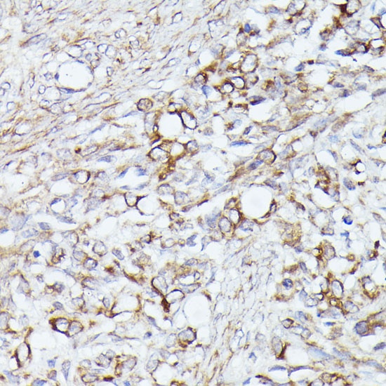
Immunohistochemistry analysis of paraffin-embedded Rat ovary using ACAT2 Rabbit pAb (CAB7866) at dilution of 1:100 (40x lens). Microwave antigen retrieval performed with 0.01M PBS Buffer (pH 7.2) prior to IHC staining.
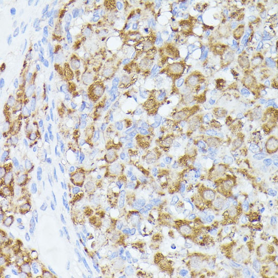
Immunohistochemistry analysis of paraffin-embedded Human oophoroma using ACAT2 Rabbit pAb (CAB7866) at dilution of 1:100 (40x lens). Microwave antigen retrieval performed with 0.01M PBS Buffer (pH 7.2) prior to IHC staining.
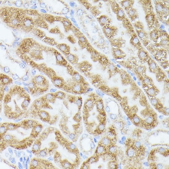
Immunohistochemistry analysis of paraffin-embedded Mouse kidney using ACAT2 Rabbit pAb (CAB7866) at dilution of 1:100 (40x lens). Microwave antigen retrieval performed with 0.01M PBS Buffer (pH 7.2) prior to IHC staining.