The p53 Antibody (CAB0263) is a high-quality antibody developed for reliable detection and analysis of target proteins. It binds specifically to the TP53 protein, allowing for precise detection and analysis in various cell types. Researchers in the fields of oncology, molecular biology, and genetics will find this antibody invaluable for their studies on TP53 and its role in cancer development and progression.
This antibody is validated for use in WB, IHC-P, IF/ICC, IP, ELISA applications and has demonstrated reactivity against Human, Mouse, Rat samples.
Product Name:
p53 Antibody
SKU:
CAB0263
Size:
20μL, 100μL
Reactivity:
Human, Mouse, Rat
Conjugate:
Unconjugated
Immunogen:
Recombinant protein (or fragment).This information is considered to be commercially sensitive.
This gene encodes a tumor suppressor protein containing transcriptional activation, DNA binding, and oligomerization domains. The encoded protein responds to diverse cellular stresses to regulate expression of target genes, thereby inducing cell cycle arrest, apoptosis, senescence, DNA repair, or changes in metabolism. Mutations in this gene are associated with a variety of human cancers, including hereditary cancers such as Li-Fraumeni syndrome. Alternative splicing of this gene and the use of alternate promoters result in multiple transcript variants and isoforms. Additional isoforms have also been shown to result from the use of alternate translation initiation codons from identical transcript variants (PMIDs: 12032546, 20937277).
Purification Method
Affinity purification
Gene ID
7157
RRID
AB_2757076
Buffer Information
Store at -20℃. Avoid freeze / thaw cycles. Buffer: PBS containing 50% glycerol, preserved with proclin300 or sodium azide, pH 7.3.
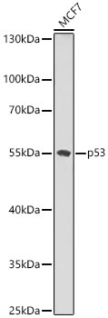
Western blot analysis of lysates from MCF7 cells using p53 Rabbit pAb (CAB0263) at 1:1000 dilution. Secondary antibody: HRP-conjugated Goat anti-Rabbit IgG (H+L) (CABS014) at 1:10000 dilution. Lysates/proteins: 25 μg per lane. Blocking buffer: 3% nonfat dry milk in TBST. Detection: ECL Basic Kit (AbGn00020). Exposure time: 30s.
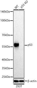
Western blot analysis of lysates from wild type (WT) and p53 knockout (KO) 293T cells using p53 Rabbit pAb (CAB0263) at 1:1000 dilution incubated overnight at 4℃. Secondary antibody: HRP-conjugated Goat anti-Rabbit IgG (H+L) (CABS014) at 1:10000 dilution. Lysates/proteins: 25 μg per lane. Blocking buffer: 3% nonfat dry milk in TBST. Detection: ECL Basic Kit (AbGn00020). Exposure time: 10s.
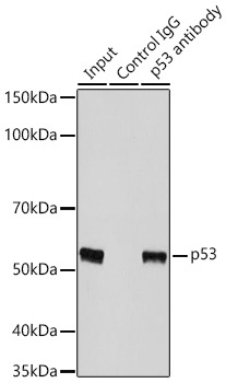
Immunoprecipitation of p53 in 200 µg extracts from 293T cells using 3 µg p53 Rabbit pAb (CAB0263). Western blot analysis was performed using p53 Rabbit pAb (CAB0263) at 1:3000 dilution.