The Ku70 Antibody (CAB0883) is a high-quality antibody developed for reliable detection and analysis of target proteins. This antibody, generated in rabbits, is highly specific for XRCC6 in human samples, making it ideal for Western blot analysis. By specifically binding to XRCC6, this antibody enables the visualization and quantification of XRCC6 levels in various cell types, facilitating research in fields such as cancer biology and genomics.XRCC6, also known as Ku70, is a critical component of the non-homologous end joining pathway for DNA double-strand break repair.
This antibody is validated for use in WB, IHC-P, IF/ICC, ELISA applications and has demonstrated reactivity against Human, Mouse, Rat samples.
Product Name:
Ku70 Antibody
SKU:
CAB0883
Size:
20μL, 100μL
Reactivity:
Human, Mouse, Rat
Conjugate:
Unconjugated
Immunogen:
Synthetic peptide. This information is considered to be commercially sensitive.
Recommended starting concentration is 1 μg/mL. Please optimize the concentration based on your specific assay requirements.
Synonyms:
ML8, KU70, TLAA, CTC75, CTCBF, G22P1, Ku70
Positive Sample:
A-549, HeLa, Hep G2, MCF7, C6
Cellular Localization:
Chromosome, Nucleus.
Calculated MW:
70kDa
Observed MW:
70kDa
The p70/p80 autoantigen is a nuclear complex consisting of two subunits with molecular masses of approximately 70 and 80 kDa. The complex functions as a single-stranded DNA-dependent ATP-dependent helicase. The complex may be involved in the repair of nonhomologous DNA ends such as that required for double-strand break repair, transposition, and V(D)J recombination. High levels of autoantibodies to p70 and p80 have been found in some patients with systemic lupus erythematosus.
Purification Method
Affinity purification
Gene ID
2547
RRID
AB_2757441
Buffer Information
Store at -20℃. Avoid freeze / thaw cycles. Buffer: PBS containing 50% glycerol, preserved with proclin300 or sodium azide, pH 7.3.
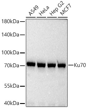
Western blot analysis of various lysates using Ku70 Rabbit pAb (CAB0883) at 1:3000 dilution incubated at room temperature for 1.5 hours. Secondary antibody: HRP-conjugated Goat anti-Rabbit IgG (H+L) (CABS014) at 1:10000 dilution. Lysates/proteins: 25 μg per lane. Blocking buffer: 3% nonfat dry milk in TBST. Detection: ECL Basic Kit (AbGn00020). Exposure time: 1s.
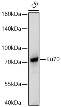
Western blot analysis of lysates from C6 cells using Ku70 Rabbit pAb (CAB0883) at 1:3000 dilution incubated at room temperature for 1.5 hours. Secondary antibody: HRP-conjugated Goat anti-Rabbit IgG (H+L) (CABS014) at 1:10000 dilution. Lysates/proteins: 25 μg per lane. Blocking buffer: 3% nonfat dry milk in TBST. Detection: ECL Basic Kit (AbGn00020). Exposure time: 45s.
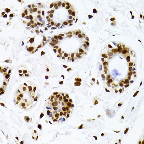
Immunohistochemistry analysis of paraffin-embedded Human breast using Ku70 Rabbit pAb (CAB0883) at dilution of 1:100 (40x lens). Microwave antigen retrieval performed with 0.01M PBS Buffer (pH 7.2) prior to IHC staining.
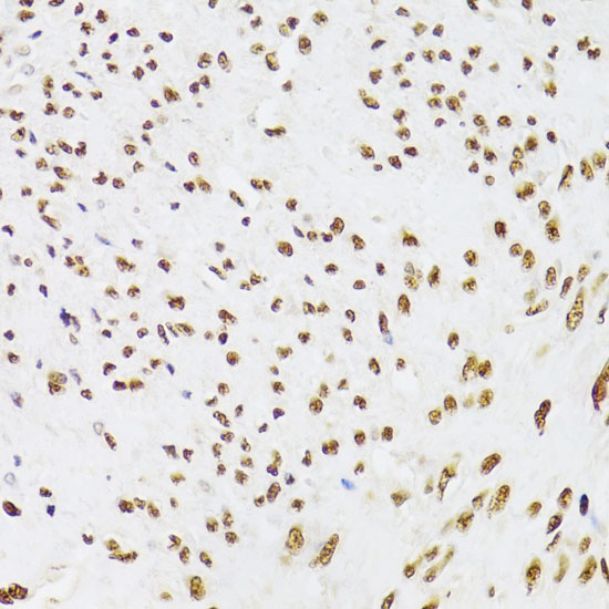
Immunohistochemistry analysis of paraffin-embedded Human leiomyoma of uterus using Ku70 Rabbit pAb (CAB0883) at dilution of 1:100 (40x lens). Microwave antigen retrieval performed with 0.01M PBS Buffer (pH 7.2) prior to IHC staining.
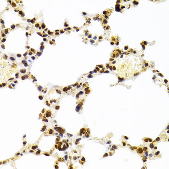
Immunohistochemistry analysis of paraffin-embedded Mouse lung using Ku70 Rabbit pAb (CAB0883) at dilution of 1:100 (40x lens). Microwave antigen retrieval performed with 0.01M PBS Buffer (pH 7.2) prior to IHC staining.
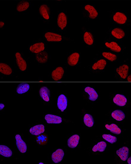
Confocal immunofluorescence analysis of U2OS cells using Ku70 Rabbit pAb (CAB0883) at dilution of 1:100. Blue: DAPI for nuclear staining.