The ACSS2 Antibody (CAB6472) is a high-quality antibody developed for reliable detection and analysis of target proteins. The antibody, produced in rabbits, is highly specific and reacts with human samples, making it suitable for use in various experimental techniques such as Western blotting.ACSS2 plays a key role in cellular metabolism, particularly in cancer cells where it is upregulated to support rapid growth and proliferation. By targeting ACSS2, researchers can gain insights into the mechanisms underlying cancer cell metabolism and potentially identify new therapeutic targets for cancer treatment.
This antibody is validated for use in WB, IF/ICC, ELISA applications and has demonstrated reactivity against Human, Mouse, Rat samples.
Product Name:
ACSS2 Antibody
SKU:
CAB6472
Size:
20μL, 100μL
Reactivity:
Human, Mouse, Rat
Conjugate:
Unconjugated
Immunogen:
Recombinant protein (or fragment).This information is considered to be commercially sensitive.
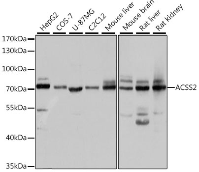
HepG2, COS-7, U-87MG, C2C12, Mouse liver, Mouse brain, Rat liver, Rat kidney
Cellular Localization:
Cytoplasm.
Calculated MW:
79kDa
Observed MW:
78kDa
This gene encodes a cytosolic enzyme that catalyzes the activation of acetate for use in lipid synthesis and energy generation. The protein acts as a monomer and produces acetyl-CoA from acetate in a reaction that requires ATP. Expression of this gene is regulated by sterol regulatory element-binding proteins, transcription factors that activate genes required for the synthesis of cholesterol and unsaturated fatty acids. Alternative splicing results in multiple transcript variants.
Purification Method
Affinity purification
Gene ID
55902
RRID
AB_2767073
Buffer Information
Store at -20℃. Avoid freeze / thaw cycles. Buffer: PBS with 0.09% Sodium azide,50% glycerol,pH7.3.
Western blot analysis of various lysates using ACSS2 Rabbit pAb (CAB6472) at 1:1000 dilution. Secondary antibody: HRP-conjugated Goat anti-Rabbit IgG (H+L) (CABS014) at 1:10000 dilution. Lysates/proteins: 25μg per lane. Blocking buffer: 3% nonfat dry milk in TBST. Detection: ECL Basic Kit (AbGn00020). Exposure time: 5s.
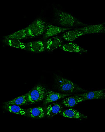
Confocal immunofluorescence analysis of NIH-3T3 cells using ACSS2 Rabbit pAb (CAB6472) at dilution of 1:200. Blue: DAPI for nuclear staining.