The ACVR1C Antibody (CAB0678) is a high-quality antibody developed for reliable detection and analysis of target proteins. This antibody, produced in rabbits, exhibits high specificity and sensitivity towards human samples and is validated for use in Western blot applications.ACVR1C, also known as Activin receptor-like kinase 7 (ALK7), plays a crucial role in various physiological processes, including bone formation, adipogenesis, and insulin sensitivity. Dysregulation of ACVR1C has been linked to diseases such as cancer, cardiovascular disorders, and metabolic syndromes.
This antibody is validated for use in WB, IHC-P, ELISA applications and has demonstrated reactivity against Human, Mouse, Rat samples.
Product Name:
ACVR1C Antibody
SKU:
CAB0678
Size:
20μL, 100μL
Reactivity:
Human, Mouse, Rat
Conjugate:
Unconjugated
Immunogen:
Recombinant protein (or fragment).This information is considered to be commercially sensitive.
Recommended starting concentration is 1 μg/mL. Please optimize the concentration based on your specific assay requirements.
Synonyms:
ALK7, ACVRLK7, ACVR1C
Positive Sample:
HT-29, BxPC-3, Mouse brain, Rat brain
Cellular Localization:
Membrane, Single-Pass Type I Membrane Protein.
Calculated MW:
55kDa
Observed MW:
55kDa
ACVR1C is a type I receptor for the TGFB (see MIM 190180) family of signaling molecules. Upon ligand binding, type I receptors phosphorylate cytoplasmic SMAD transcription factors, which then translocate to the nucleus and interact directly with DNA or in complex with other transcription factors (Bondestam et al., 2001 [PubMed 12063393]).
Purification Method
Affinity purification
Gene ID
130399
RRID
AB_2757330
Buffer Information
Store at -20℃. Avoid freeze / thaw cycles. Buffer: PBS containing 50% glycerol, preserved with proclin300 or sodium azide, pH 7.3.
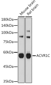
Western blot analysis of various lysates using ACVR1C Rabbit pAb (CAB0678) at 1:1000 dilution. Secondary antibody: HRP-conjugated Goat anti-Rabbit IgG (H+L) (CABS014) at 1:10000 dilution. Lysates/proteins: 25μg per lane. Blocking buffer: 3% nonfat dry milk in TBST. Detection: ECL Basic Kit (AbGn00020). Exposure time: 180s.
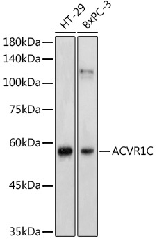
Western blot analysis of various lysates using ACVR1C Rabbit pAb (CAB0678) at 1:1000 dilution. Secondary antibody: HRP-conjugated Goat anti-Rabbit IgG (H+L) (CABS014) at 1:10000 dilution. Lysates/proteins: 25μg per lane. Blocking buffer: 3% nonfat dry milk in TBST. Detection: ECL Enhanced Kit (AbGn00021). Exposure time: 180s.
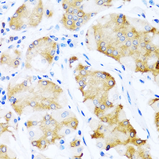
Immunohistochemistry analysis of paraffin-embedded Human stomach using ACVR1C Rabbit pAb (CAB0678) at dilution of 1:100 (40x lens). Microwave antigen retrieval performed with 0.01M PBS Buffer (pH 7.2) prior to IHC staining.
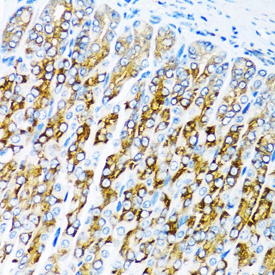
Immunohistochemistry analysis of paraffin-embedded Mouse stomach using ACVR1C Rabbit pAb (CAB0678) at dilution of 1:100 (40x lens). Microwave antigen retrieval performed with 0.01M PBS Buffer (pH 7.2) prior to IHC staining.