The ADCYAP1R1 Antibody (CAB3120) is a high-quality antibody developed for reliable detection and analysis of target proteins. This antibody, generated in rabbits, exhibits high specificity and sensitivity for human samples, making it suitable for use in Western blot applications.ADCYAP1R1, also known as PAC1 receptor, plays a crucial role in mediating the effects of pituitary adenylate cyclase-activating polypeptide (PACAP), a neuropeptide involved in the regulation of stress response, neurotransmission, and immune functions. The ADCYAP1R1 Polyclonal Antibody enables researchers to detect and analyze the expression of ADCYAP1R1 in different cell types, providing insights into its role in various physiological and pathological conditions.
This antibody is validated for use in WB, IF/ICC, ELISA applications and has demonstrated reactivity against Human, Mouse, Rat samples.
Product Name:
ADCYAP1R1 Antibody
SKU:
CAB3120
Size:
20μL, 100μL
Reactivity:
Human, Mouse, Rat
Conjugate:
Unconjugated
Immunogen:
Recombinant protein (or fragment).This information is considered to be commercially sensitive.
Recommended starting concentration is 1 μg/mL. Please optimize the concentration based on your specific assay requirements.
Synonyms:
PAC1, PAC1R, PACAPR, PACAPRI, ADCYAP1R1
Positive Sample:
U-87MG, Mouse heart
Cellular Localization:
Cell Membrane, Multi-Pass Membrane Protein.
Calculated MW:
53kDa
Observed MW:
53kDa
This gene encodes type I adenylate cyclase activating polypeptide receptor, which is a membrane-associated protein and shares significant homology with members of the glucagon/secretin receptor family. This receptor mediates diverse biological actions of adenylate cyclase activating polypeptide 1 and is positively coupled to adenylate cyclase. Multiple alternatively spliced transcript variants encoding distinct isoforms have been identified.
Purification Method
Affinity purification
Gene ID
117
RRID
AB_2764918
Buffer Information
Store at -20℃. Avoid freeze / thaw cycles. Buffer: PBS containing 50% glycerol, preserved with proclin300 or sodium azide, pH 7.3.
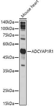
Western blot analysis of lysates from Mouse heart , using ADCYAP1R1 Rabbit pAb (CAB3120) at 1:1000 dilution. Secondary antibody: HRP-conjugated Goat anti-Rabbit IgG (H+L) (CABS014) at 1:10000 dilution. Lysates/proteins: 25μg per lane. Blocking buffer: 3% nonfat dry milk in TBST. Detection: ECL Basic Kit (AbGn00020). Exposure time: 5s.
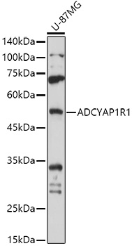
Western blot analysis of lysates from U-87MG cells, using ADCYAP1R1 Rabbit pAb (CAB3120) at 1:1000 dilution. Secondary antibody: HRP-conjugated Goat anti-Rabbit IgG (H+L) (CABS014) at 1:10000 dilution. Lysates/proteins: 25μg per lane. Blocking buffer: 3% nonfat dry milk in TBST. Detection: ECL Basic Kit (AbGn00020). Exposure time: 30s.
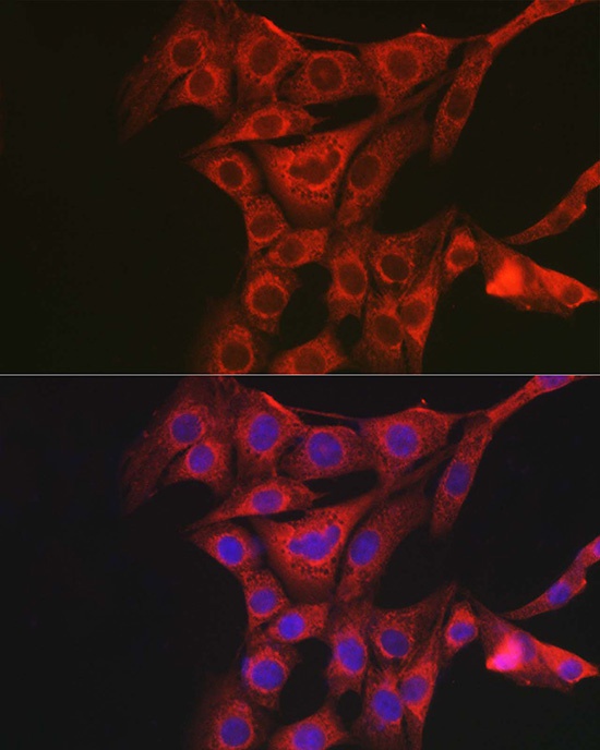
Immunofluorescence analysis of NIH/3T3 cells using ADCYAP1R1 Rabbit pAb (CAB3120) at dilution of 1:100 (40x lens). Secondary antibody: Cy3-conjugated Goat anti-Rabbit IgG (H+L) (CABS007) at 1:500 dilution. Blue: DAPI for nuclear staining.
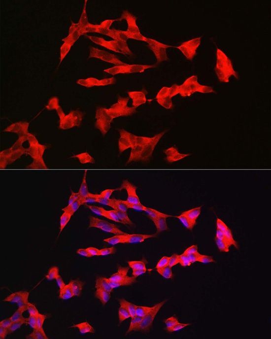
Immunofluorescence analysis of SH-SY5Y cells using ADCYAP1R1 Rabbit pAb (CAB3120) at dilution of 1:100 (40x lens). Secondary antibody: Cy3-conjugated Goat anti-Rabbit IgG (H+L) (CABS007) at 1:500 dilution. Blue: DAPI for nuclear staining.