The ADK Antibody (CAB15023) is a high-quality antibody developed for reliable detection and analysis of target proteins. ADK is known to play an important role in purine metabolism and is implicated in various biological processes, including inflammation, immune response, and neurotransmission.This antibody, produced in rabbits, is highly specific to human ADK and has been validated for use in Western blot applications. Its binding affinity to ADK allows for accurate detection and quantification of the enzyme in different experimental settings, making it an essential tool for studies in biochemistry, pharmacology, and neuroscience.The ADK Polyclonal Antibody is particularly useful for investigating the role of ADK in various diseases, such as cancer, neurodegenerative disorders, and autoimmune conditions.
This antibody is validated for use in WB, IHC-P, IF/ICC, ELISA applications and has demonstrated reactivity against Human, Mouse, Rat samples.
Product Name:
ADK Antibody
SKU:
CAB15023
Size:
20μL, 100μL
Reactivity:
Human, Mouse, Rat
Conjugate:
Unconjugated
Immunogen:
Recombinant protein (or fragment).This information is considered to be commercially sensitive.
Recommended starting concentration is 1 μg/mL. Please optimize the concentration based on your specific assay requirements.
Synonyms:
AK, ADK
Positive Sample:
LO2, A-431, Mouse testis, Mouse kidney, Mouse spleen, Rat liver
Cellular Localization:
Cytoplasm, Nucleus.
Calculated MW:
41kDa
Observed MW:
41kDa
This gene an enzyme which catalyzes the transfer of the gamma-phosphate from ATP to adenosine, thereby serving as a regulator of concentrations of both extracellular adenosine and intracellular adenine nucleotides. Adenosine has widespread effects on the cardiovascular, nervous, respiratory, and immune systems and inhibitors of the enzyme could play an important pharmacological role in increasing intravascular adenosine concentrations and acting as anti-inflammatory agents. Multiple transcript variants encoding different isoforms have been found for this gene.
Purification Method
Affinity purification
Gene ID
132
RRID
AB_2761903
Buffer Information
Store at -20℃. Avoid freeze / thaw cycles. Buffer: PBS with 0.01% thimerosal,50% glycerol,pH7.3.
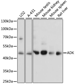
Western blot analysis of various lysates using ADK Rabbit pAb (CAB15023) at 1:1000 dilution. Secondary antibody: HRP-conjugated Goat anti-Rabbit IgG (H+L) (CABS014) at 1:10000 dilution. Lysates/proteins: 25μg per lane. Blocking buffer: 3% nonfat dry milk in TBST. Detection: ECL Basic Kit (AbGn00020). Exposure time: 5s.
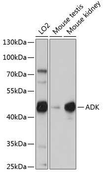
Western blot analysis of various lysates using ADK Rabbit pAb (CAB15023) at 1:1000 dilution. Secondary antibody: HRP-conjugated Goat anti-Rabbit IgG (H+L) (CABS014) at 1:10000 dilution. Lysates/proteins: 25μg per lane. Blocking buffer: 3% nonfat dry milk in TBST. Detection: ECL Basic Kit (AbGn00020). Exposure time: 90s.
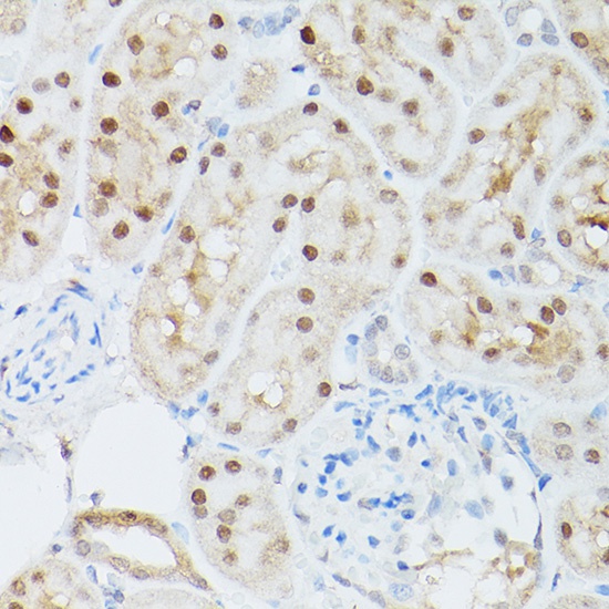
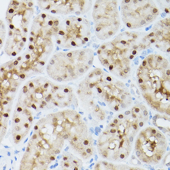
Immunohistochemistry analysis of paraffin-embedded Mouse kidney using ADK Rabbit pAb (CAB15023) at dilution of 1:300 (40x lens). High pressure antigen retrieval performed with 0.01M Citrate buffer (pH 6.0) prior to IHC staining.
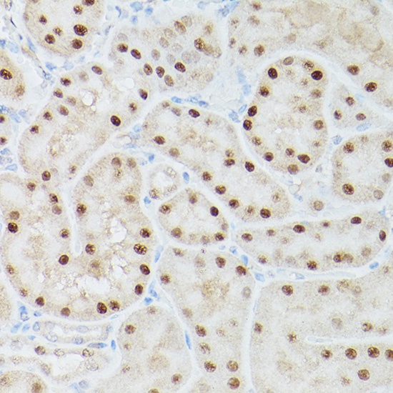
Immunohistochemistry analysis of paraffin-embedded Rat kidney using ADK Rabbit pAb (CAB15023) at dilution of 1:300 (40x lens). High pressure antigen retrieval performed with 0.01M Citrate buffer (pH 6.0) prior to IHC staining.
Immunohistochemistry analysis of paraffin-embedded Mouse kidney using ADK Rabbit pAb (CAB15023) at dilution of 1:300 (40x lens). High pressure antigen retrieval performed with 0.01M Citrate buffer (pH 6.0) prior to IHC staining.
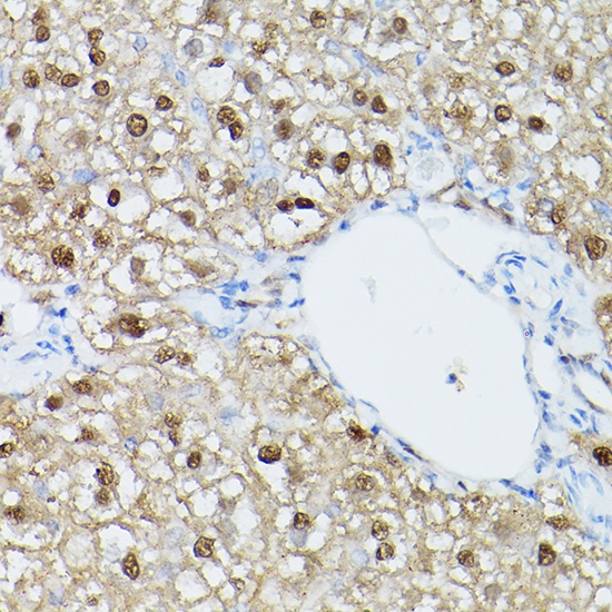
Immunohistochemistry analysis of paraffin-embedded Rat liver using ADK Rabbit pAb (CAB15023) at dilution of 1:300 (40x lens). High pressure antigen retrieval performed with 0.01M Citrate buffer (pH 6.0) prior to IHC staining.
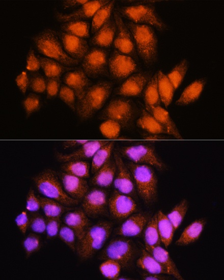
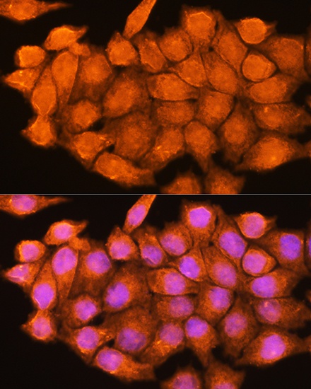
Immunofluorescence analysis of HeLa cells using ADK Rabbit pAb (CAB15023) at dilution of 1:100. Secondary antibody: Cy3-conjugated Goat anti-Rabbit IgG (H+L) (CABS007) at 1:500 dilution. Blue: DAPI for nuclear staining.
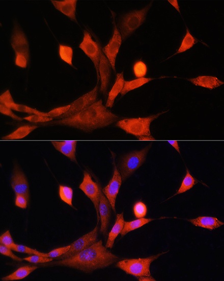
Immunofluorescence analysis of NIH/3T3 cells using ADK Rabbit pAb (CAB15023) at dilution of 1:100. Secondary antibody: Cy3-conjugated Goat anti-Rabbit IgG (H+L) (CABS007) at 1:500 dilution. Blue: DAPI for nuclear staining.
Immunofluorescence analysis of HeLa cells using ADK Rabbit pAb (CAB15023) at dilution of 1:100. Secondary antibody: Cy3-conjugated Goat anti-Rabbit IgG (H+L) (CABS007) at 1:500 dilution. Blue: DAPI for nuclear staining.