The ADORA2B Antibody (CAB1953) is a high-quality antibody developed for reliable detection and analysis of target proteins. This antibody, produced in rabbits, has been rigorously validated for use in Western blot applications and exhibits high reactivity with human samples.Adora2b, also known as the adenosine A2B receptor, is a G protein-coupled receptor that plays a key role in regulating adenosine-mediated signaling pathways. Its involvement in modulating immune responses, inflammation, and tissue repair makes it a promising target for therapeutic interventions in conditions such as cardiovascular disease, cancer, and inflammatory disorders.
This antibody is validated for use in WB, IF/ICC, ELISA applications and has demonstrated reactivity against Mouse, Rat samples.
Product Name:
ADORA2B Antibody
SKU:
CAB1953
Size:
20μL, 100μL
Reactivity:
Mouse, Rat
Conjugate:
Unconjugated
Immunogen:
Synthetic peptide. This information is considered to be commercially sensitive.
Recommended starting concentration is 1 μg/mL. Please optimize the concentration based on your specific assay requirements.
Synonyms:
ADORA2, ADORA2B
Positive Sample:
A-431, 293T, Mouse brain, Rat testis
Cellular Localization:
Cell Membrane, Multi-Pass Membrane Protein.
Calculated MW:
36kDa
Observed MW:
36kDa/30-50kDa
This gene encodes an adenosine receptor that is a member of the G protein-coupled receptor superfamily. This integral membrane protein stimulates adenylate cyclase activity in the presence of adenosine. This protein also interacts with netrin-1, which is involved in axon elongation. The gene is located near the Smith-Magenis syndrome region on chromosome 17.
Purification Method
Affinity purification
Gene ID
136
RRID
AB_2763979
Buffer Information
Store at -20℃. Avoid freeze / thaw cycles. Buffer: PBS containing 50% glycerol, preserved with proclin300 or sodium azide, pH 7.3.
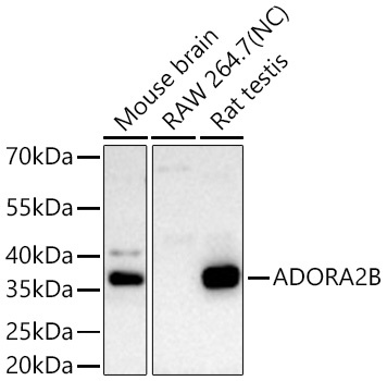
Western blot analysis of various lysates using ADORA2B Rabbit pAb (CAB1953) at 1:1000 dilution incubated overnight at 4℃. Secondary antibody: HRP-conjugated Goat anti-Rabbit IgG (H+L) (CABS014) at 1:10000 dilution. Lysates/proteins: 25 μg per lane. Blocking buffer: 3% nonfat dry milk in TBST. Detection: ECL Basic Kit (AbGn00020). Negative control (NC): RAW 264.7. Exposure time: 90 s.
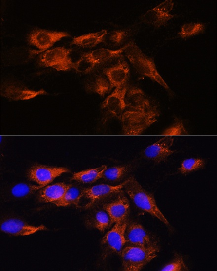
Immunofluorescence analysis of C6 cells using ADORA2B Rabbit pAb (CAB1953) at dilution of 1:100. Secondary antibody: Cy3-conjugated Goat anti-Rabbit IgG (H+L) (CABS007) at 1:500 dilution. Blue: DAPI for nuclear staining.