The AGR2 Monoclonal Antibody (CAB12411) is a high-quality antibody developed for reliable detection and analysis of target proteins. This antibody, produced using innovative rabbit monoclonal technology, boasts high specificity and sensitivity for detecting AGR2 in human samples, making it a valuable asset in cancer research.AGR2, also known as anterior gradient protein 2 homolog, is involved in various cellular processes, including cell growth, differentiation, and metastasis. Its overexpression has been linked to several types of cancer, making it a potential biomarker for early cancer detection and a target for therapeutic interventions.
This antibody is validated for use in WB, IF/ICC, ELISA applications and has demonstrated reactivity against Human, Mouse samples.
Product Name:
AGR2 Monoclonal Antibody
SKU:
CAB12411
Size:
20μL, 100μL
Reactivity:
Human, Mouse
Clone Number:
ARC0709
Conjugate:
Unconjugated
Immunogen:
Synthetic peptide. This information is considered to be commercially sensitive.
This gene encodes a member of the disulfide isomerase (PDI) family of endoplasmic reticulum (ER) proteins that catalyze protein folding and thiol-disulfide interchange reactions. The encoded protein has an N-terminal ER-signal sequence, a catalytically active thioredoxin domain, and a C-terminal ER-retention sequence. This protein plays a role in cell migration, cellular transformation and metastasis and is as a p53 inhibitor. As an ER-localized molecular chaperone, it plays a role in the folding, trafficking, and assembly of cysteine-rich transmembrane receptors and the cysteine-rich intestinal gylcoprotein mucin. This gene has been implicated in inflammatory bowel disease and cancer progression.
Purification Method
Affinity purification
Gene ID
10551
RRID
AB_2861663
Buffer Information
Store at -20℃. Avoid freeze / thaw cycles. Buffer: PBS containing 50% glycerol and 0.05% BSA, preserved with proclin300 or sodium azide, pH 7.3.
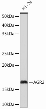
Western blot analysis of lysates from HT-29 cells, using AGR2 Rabbit mAb (CAB12411) at 1:1000 dilution. Secondary antibody: HRP-conjugated Goat anti-Rabbit IgG (H+L) (CABS014) at 1:10000 dilution. Lysates/proteins: 25μg per lane. Blocking buffer: 3% nonfat dry milk in TBST. Detection: ECL Basic Kit (AbGn00020). Exposure time: 10s.
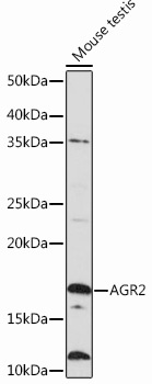
Western blot analysis of lysates from Mouse testis, using AGR2 Rabbit mAb (CAB12411) at 1:1000 dilution. Secondary antibody: HRP-conjugated Goat anti-Rabbit IgG (H+L) (CABS014) at 1:10000 dilution. Lysates/proteins: 25μg per lane. Blocking buffer: 3% nonfat dry milk in TBST. Detection: ECL Basic Kit (AbGn00020). Exposure time: 3min.