The AHCY Antibody (CAB5300) is a high-quality antibody developed for reliable detection and analysis of target proteins. This antibody, generated in rabbits, is highly specific to human AHCY and has been validated for use in Western blot applications. It detects the AHCY protein, allowing for precise analysis in various cell types, making it an ideal choice for studies in biochemistry and molecular biology.
This antibody is validated for use in WB, IF/ICC, ELISA applications and has demonstrated reactivity against Human, Mouse, Rat samples.
Product Name:
AHCY Antibody
SKU:
CAB5300
Size:
20μL, 100μL
Reactivity:
Human, Mouse, Rat
Conjugate:
Unconjugated
Immunogen:
Recombinant protein (or fragment).This information is considered to be commercially sensitive.
Recommended starting concentration is 1 μg/mL. Please optimize the concentration based on your specific assay requirements.
Synonyms:
SAHH, adoHcyase, AHCY
Positive Sample:
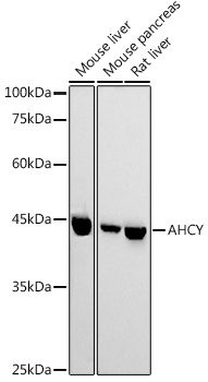
Mouse liver, Mouse pancreas, Rat liver
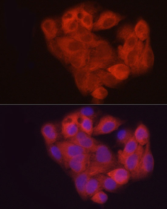
Cellular Localization:
Cytoplasm, Melanosome.
Calculated MW:
48kDa
Observed MW:
45kDa
S-adenosylhomocysteine hydrolase belongs to the adenosylhomocysteinase family. It catalyzes the reversible hydrolysis of S-adenosylhomocysteine (AdoHcy) to adenosine (Ado) and L-homocysteine (Hcy). Thus, it regulates the intracellular S-adenosylhomocysteine (SAH) concentration thought to be important for transmethylation reactions. Deficiency in this protein is one of the different causes of hypermethioninemia. Alternatively spliced transcript variants encoding different isoforms have been found for this gene.
Purification Method
Affinity purification
Gene ID
191
RRID
AB_2766112
Buffer Information
Store at -20℃. Avoid freeze / thaw cycles. Buffer: PBS containing 50% glycerol, preserved with proclin300 or sodium azide, pH 7.3.
Western blot analysis of various lysates using AHCY Rabbit pAb (CAB5300) at 1:1000 dilution. Secondary antibody: HRP-conjugated Goat anti-Rabbit IgG (H+L) (CABS014) at 1:10000 dilution. Lysates/proteins: 25μg per lane. Blocking buffer: 3% nonfat dry milk in TBST. Detection: ECL Basic Kit (AbGn00020). Exposure time: 180s.
Immunofluorescence analysis of HepG2 cells using AHCY Rabbit pAb (CAB5300) at dilution of 1:50 (40x lens). Secondary antibody: Cy3-conjugated Goat anti-Rabbit IgG (H+L) (CABS007) at 1:500 dilution. Blue: DAPI for nuclear staining.