The AIP Antibody (CAB12546) is a high-quality antibody developed for reliable detection and analysis of target proteins. This antibody, produced in rabbits, exhibits high reactivity with human samples and is validated for use in Western blot applications. By targeting the AIP protein, this antibody enables precise detection and analysis in a variety of cell types, making it an essential component for studies in immunology and cancer research.AIP, also known as aryl hydrocarbon receptor-interacting protein, is involved in controlling immune responses and has been implicated in various diseases such as cancer, autoimmune disorders, and chronic inflammatory conditions.
This antibody is validated for use in WB, ELISA applications and has demonstrated reactivity against Human, Mouse, Rat samples.
Product Name:
AIP Antibody
SKU:
CAB12546
Size:
20μL, 100μL
Reactivity:
Human, Mouse, Rat
Conjugate:
Unconjugated
Immunogen:
Recombinant protein (or fragment).This information is considered to be commercially sensitive.
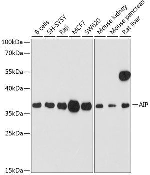
B cells, SH-SY5Y, Raji, MCF7, SW620, Mouse kidney, Mouse pancreas, Rat liver
Cellular Localization:
Cytoplasm.
Calculated MW:
38kDa
Observed MW:
37kDa
The protein encoded by this gene is a receptor for aryl hydrocarbons and a ligand-activated transcription factor. The encoded protein is found in the cytoplasm as part of a multiprotein complex, but upon binding of ligand is transported to the nucleus. This protein can regulate the expression of many xenobiotic metabolizing enzymes. Also, the encoded protein can bind specifically to and inhibit the activity of hepatitis B virus. Three transcript variants encoding different isoforms have been found for this gene.
Purification Method
Affinity purification
Gene ID
9049
RRID
AB_2759386
Buffer Information
Store at -20℃. Avoid freeze / thaw cycles. Buffer: PBS containing 50% glycerol, preserved with proclin300 or sodium azide, pH 7.3.
Western blot analysis of various lysates using AIP Rabbit pAb (CAB12546) at 1:1000 dilution. Secondary antibody: HRP-conjugated Goat anti-Rabbit IgG (H+L) (CABS014) at 1:10000 dilution. Lysates/proteins: 25μg per lane. Blocking buffer: 3% nonfat dry milk in TBST. Detection: ECL Enhanced Kit (AbGn00021). Exposure time: 90s.
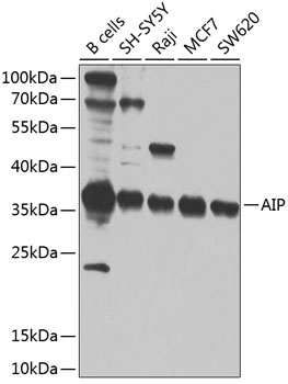
Western blot analysis of various lysates using AIP Rabbit pAb (CAB12546) at 1:1000 dilution. Secondary antibody: HRP-conjugated Goat anti-Rabbit IgG (H+L) (CABS014) at 1:10000 dilution. Lysates/proteins: 25μg per lane. Blocking buffer: 3% nonfat dry milk in TBST. Detection: ECL Enhanced Kit (AbGn00021). Exposure time: 90s.