The AKAP8 Antibody (CAB17302) is a high-quality antibody developed for reliable detection and analysis of target proteins. This antibody, generated in rabbits, is specifically designed to target AKAP8 in human samples, making it an essential tool for Western blot applications.AKAP8, also known as A-kinase anchor protein 8, is a key player in cellular signaling pathways, particularly those involving protein kinase A (PKA) and other signaling molecules.
This antibody is validated for use in WB, IHC-P, ELISA applications and has demonstrated reactivity against Human, Rat samples.
Product Name:
AKAP8 Antibody
SKU:
CAB17302
Size:
20μL, 100μL
Reactivity:
Human, Rat
Conjugate:
Unconjugated
Immunogen:
Synthetic peptide. This information is considered to be commercially sensitive.
Recommended starting concentration is 1 μg/mL. Please optimize the concentration based on your specific assay requirements.
Synonyms:
AKAP-8, AKAP95, AKAP 95, AKAP-95, AKAP8
Positive Sample:
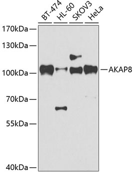
BT-474, HL-60, SKOV3, HeLa
Cellular Localization:
Nucleus Matrix.
Calculated MW:
76kDa
Observed MW:
100kDa
This gene encodes a member of the A-kinase anchor protein family. A-kinase anchor proteins are scaffold proteins that contain a binding domain for the RI/RII subunit of protein kinase A (PKA) and recruit PKA and other signaling molecules to specific subcellular locations. This gene encodes a nuclear A-kinase anchor protein that binds to the RII alpha subunit of PKA and may play a role in chromosome condensation during mitosis by targeting PKA and the condensin complex to chromatin. A pseudogene of this gene is located on the short arm of chromosome 9.
Purification Method
Affinity purification
Gene ID
10270
RRID
AB_2768304
Buffer Information
Store at -20℃. Avoid freeze / thaw cycles. Buffer: PBS containing 50% glycerol, preserved with proclin300 or sodium azide, pH 7.3.
Western blot analysis of various lysates using AKAP8 Rabbit pAb (CAB17302) at 1:500 dilution. Secondary antibody: HRP-conjugated Goat anti-Rabbit IgG (H+L) (CABS014) at 1:10000 dilution. Lysates/proteins: 25μg per lane. Blocking buffer: 3% nonfat dry milk in TBST. Detection: ECL Basic Kit (AbGn00020). Exposure time: 90s.
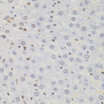
Immunohistochemistry analysis of paraffin-embedded Rat liver using AKAP8 Rabbit pAb (CAB17302) (40x lens). Microwave antigen retrieval performed with 0.01M PBS Buffer (pH 7.2) prior to IHC staining.