The AKT1S1/PRAS40 Polyclonal Antibody (CAB6238) is a high-quality antibody developed for reliable detection and analysis of target proteins. This antibody, produced in rabbits, exhibits high reactivity with human samples and has been validated for use in Western blot applications.The AKT1S1/PRAS40 protein is a critical player in the Akt/mTOR pathway, impacting cell proliferation and survival through its interactions with various downstream effectors. Dysregulation of this pathway has been implicated in numerous diseases, including cancer, making the AKT1S1/PRAS40 protein a promising target for therapeutic interventions.
This antibody is validated for use in WB, IHC-P, IF/ICC, ELISA applications and has demonstrated reactivity against Human, Mouse, Rat samples.
Product Name:
AKT1S1/PRAS40 Polyclonal Antibody
SKU:
CAB6238
Size:
20μL, 100μL
Reactivity:
Human, Mouse, Rat
Conjugate:
Unconjugated
Immunogen:
Synthetic peptide. This information is considered to be commercially sensitive.
Recommended starting concentration is 1 μg/mL. Please optimize the concentration based on your specific assay requirements.
Synonyms:
Lobe, PRAS40, AKT1S1/PRAS40
Positive Sample:
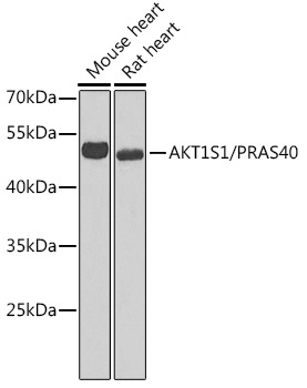
Mouse heart, Rat heart
Cellular Localization:
Cytoplasm, Cytosol.
Calculated MW:
27kDa
Observed MW:
50kDa
AKT1S1 is a proline-rich substrate of AKT (MIM 164730) that binds 14-3-3 protein (see YWHAH, MIM 113508) when phosphorylated (Kovacina et al., 2003 [PubMed 12524439]).
Purification Method
Affinity purification
Gene ID
84335
RRID
AB_2766846
Buffer Information
Store at -20℃. Avoid freeze / thaw cycles. Buffer: PBS containing 50% glycerol, preserved with proclin300 or sodium azide, pH 7.3.
Western blot analysis of various lysates using AKT1S1/PRAS40 Rabbit pAb (CAB6238) at 1:1000 dilution. Secondary antibody: HRP-conjugated Goat anti-Rabbit IgG (H+L) (CABS014) at 1:10000 dilution. Lysates/proteins: 25μg per lane. Blocking buffer: 3% nonfat dry milk in TBST. Detection: ECL Enhanced Kit (AbGn00021). Exposure time: 90s.
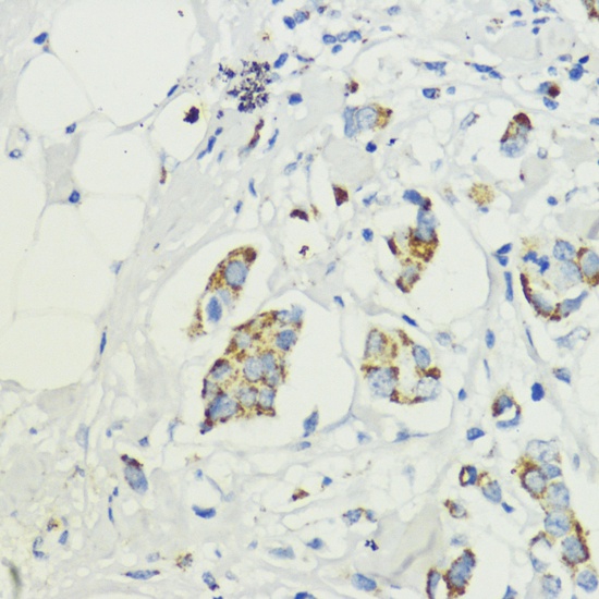
Immunohistochemistry analysis of paraffin-embedded Human mammary cancer using AKT1S1/PRAS40 Rabbit pAb (CAB6238) at dilution of 1:200 (40x lens). Microwave antigen retrieval performed with 0.01M PBS Buffer (pH 7.2) prior to IHC staining.
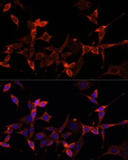
Immunofluorescence analysis of NIH-3T3 cells using AKT1S1/PRAS40 Rabbit pAb (CAB6238) at dilution of 1:100 (40x lens). Secondary antibody: Cy3-conjugated Goat anti-Rabbit IgG (H+L) (CABS007) at 1:500 dilution. Blue: DAPI for nuclear staining.
 at 1:1000 dilution. Secondary antibody: HRP Goat Anti-Rabbit IgG (H+L) at 1:10000 dilution. Lysates/proteins: 25μg per lane. Blocking buffer: 3% nonfat dry milk in TBST.")
 at 1:1000 dilution. Secondary antibody: HRP Goat Anti-Rabbit IgG (H+L) at 1:10000 dilution. Lysates/proteins: 25μg per lane. Blocking buffer: 3% nonfat dry milk in TBST.")
")


