The ALDH3A1 Antibody (CAB5502) is a high-quality antibody developed for reliable detection and analysis of target proteins. Produced in rabbits, this antibody is highly specific to human ALDH3A1 and has been validated for Western blot applications. By binding to the ALDH3A1 protein, this antibody enables the detection and analysis of ALDH3A1 in various cell types, making it ideal for studies in biology, biochemistry, and toxicology.ALDH3A1 is highly expressed in tissues that are exposed to environmental toxins, such as the liver and lung, where it plays a crucial role in protecting cells from oxidative stress.
This antibody is validated for use in WB, IF/ICC, ELISA, IF-P applications and has demonstrated reactivity against Human, Mouse, Rat samples.
Product Name:
ALDH3A1 Antibody
SKU:
CAB5502
Size:
20μL, 100μL
Reactivity:
Human, Mouse, Rat
Conjugate:
Unconjugated
Immunogen:
Recombinant protein (or fragment).This information is considered to be commercially sensitive.
Recommended starting concentration is 1 μg/mL. Please optimize the concentration based on your specific assay requirements.
Synonyms:
ALDH3, ALDHIII, ALDH3A1
Positive Sample:
A-549
Cellular Localization:
Cytoplasm.
Calculated MW:
50kDa
Observed MW:
55kDa
Aldehyde dehydrogenases oxidize various aldehydes to the corresponding acids. They are involved in the detoxification of alcohol-derived acetaldehyde and in the metabolism of corticosteroids, biogenic amines, neurotransmitters, and lipid peroxidation. The enzyme encoded by this gene forms a cytoplasmic homodimer that preferentially oxidizes aromatic and medium-chain (6 carbons or more) saturated and unsaturated aldehyde substrates. It is thought to promote resistance to UV and 4-hydroxy-2-nonenal-induced oxidative damage in the cornea. The gene is located within the Smith-Magenis syndrome region on chromosome 17. Multiple alternatively spliced variants, encoding the same protein, have been identified.
Purification Method
Affinity purification
Gene ID
218
RRID
AB_2766298
Buffer Information
Store at -20℃. Avoid freeze / thaw cycles. Buffer: PBS containing 50% glycerol, preserved with proclin300 or sodium azide, pH 7.3.
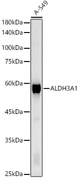
Western blot analysis of lysates from A-549 cells, using ALDH3A1 Rabbit pAb (CAB5502) at 1:700 dilution. Secondary antibody: HRP-conjugated Goat anti-Rabbit IgG (H+L) (CABS014) at 1:10000 dilution. Lysates/proteins: 25μg per lane. Blocking buffer: 3% nonfat dry milk in TBST. Detection: ECL Basic Kit (AbGn00020). Exposure time: 5s.
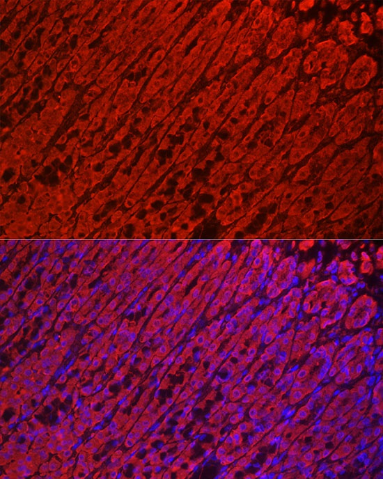
Immunofluorescence analysis of paraffin-embedded Rat stomach using ALDH3A1 Rabbit pAb (CAB5502) at dilution of 1:100 (40x lens). Secondary antibody: Cy3-conjugated Goat anti-Rabbit IgG (H+L) (CABS007) at 1:500 dilution. Blue: DAPI for nuclear staining.
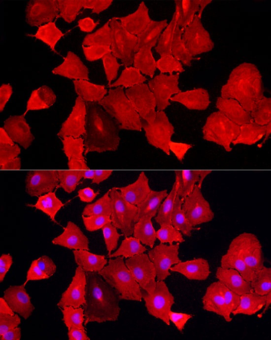
Immunofluorescence analysis of A-549 cells using ALDH3A1 Rabbit pAb (CAB5502) at dilution of 1:100 (40x lens). Secondary antibody: Cy3-conjugated Goat anti-Rabbit IgG (H+L) (CABS007) at 1:500 dilution. Blue: DAPI for nuclear staining.
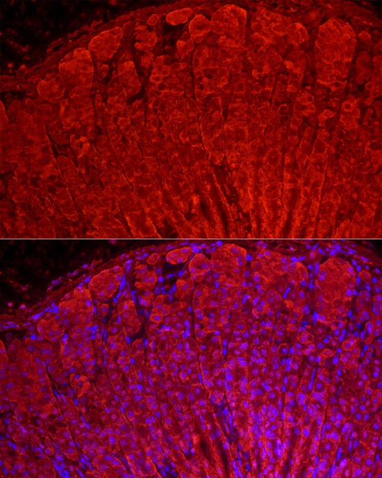
Immunofluorescence analysis of paraffin-embedded Mouse stomach using ALDH3A1 Rabbit pAb (CAB5502) at dilution of 1:100 (40x lens). Secondary antibody: Cy3-conjugated Goat anti-Rabbit IgG (H+L) (CABS007) at 1:500 dilution. Blue: DAPI for nuclear staining.