The AP2A1 Monoclonal Antibody (CAB4403) is a high-quality antibody developed for reliable detection and analysis of target proteins. This gene encodes the alpha 1 adaptin subunit of the adaptor protein 2 (AP-2) complex found in clathrin coated vesicles. The AP-2 complex is a heterotetramer consisting of two large adaptins (alpha or beta), a medium adaptin (mu), and a small adaptin (sigma). The complex is part of the protein coat on the cytoplasmic face of coated vesicles which links clathrin to receptors in vesicles. Alternative splicing of this gene results in two transcript variants encoding two different isoforms. A third transcript variant has been described, but its full length nature has not been determined.
This antibody is validated for use in WB, IF/ICC, ELISA applications and has demonstrated reactivity against Human, Mouse, Rat samples.
Product Name:
AP2A1 Monoclonal Antibody
SKU:
CAB4403
Size:
100μL, 20μL
Reactivity:
Human, Mouse, Rat
Clone Number:
ARC0998
Conjugate:
Unconjugated
Immunogen:
Recombinant protein (or fragment).This information is considered to be commercially sensitive.
Tested Applications:
WBIF/ICCELISA
Recommended Dilution:
WB
1:1000 - 1:2000
IF/ICC
1:50 - 1:200
ELISA
Recommended starting concentration is 1 μg/mL. Please optimize the concentration based on your specific assay requirements.
Synonyms:
ADTAA, CLAPA1, AP2-ALPHA, AP2A1
Positive Sample:
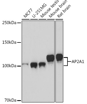
MCF7, U-251MG, Mouse testis, Mouse brain, Rat brain
This gene encodes the alpha 1 adaptin subunit of the adaptor protein 2 (AP-2) complex found in clathrin coated vesicles. The AP-2 complex is a heterotetramer consisting of two large adaptins (alpha or beta), a medium adaptin (mu), and a small adaptin (sigma). The complex is part of the protein coat on the cytoplasmic face of coated vesicles which links clathrin to receptors in vesicles. Alternative splicing of this gene results in two transcript variants encoding two different isoforms. A third transcript variant has been described, but its full length nature has not been determined.
Purification Method
Affinity purification
Gene ID
160
RRID
AB_2863264
Buffer Information
Store at -20℃. Avoid freeze / thaw cycles. Buffer: PBS containing 50% glycerol and 0.05% BSA, preserved with proclin300 or sodium azide, pH 7.3.
Western blot analysis of various lysates using AP2A1 Rabbit mAb (CAB4403) at 1:1000 dilution. Secondary antibody: HRP-conjugated Goat anti-Rabbit IgG (H+L) (AS014) at 1:10000 dilution. Lysates/proteins: 25μg per lane. Blocking buffer: 3% nonfat dry milk in TBST. Detection: ECL Basic Kit (AbGn00020). Exposure time: 90s.
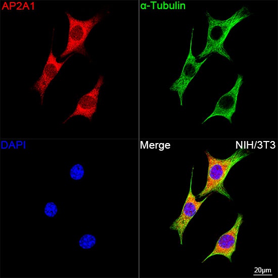
Confocal imaging of NIH/3T3 cells using AP2A1 Rabbit mAb (CAB4403, dilution 1:100) followed by a further incubation with Cy3 Goat Anti-Rabbit IgG (H+L) (AS007, dilution 1:500) (Red). The cells were counterstained with α-Tubulin Mouse mAb (AC012, dilution 1:400) followed by incubation with ABflo® 488-conjugated Goat Anti-Mouse IgG (H+L) Ab (AS076, dilution 1:500) (Green). DAPI was used for nuclear staining (Blue). Objective: 100x.