The Alkaline Phosphatase (ALPL) Antibody (CAB1080) is a high-quality antibody developed for reliable detection and analysis of target proteins. This gene encodes a member of the alkaline phosphatase family of proteins. There are at least four distinct but related alkaline phosphatases: intestinal, placental, placental-like, and liver/bone/kidney (tissue non-specific). The first three are located together on chromosome 2, while the tissue non-specific form is located on chromosome 1. The product of this gene is a membrane bound glycosylated enzyme that is not expressed in any particular tissue and is, therefore, referred to as the tissue-nonspecific form of the enzyme. Alternative splicing results in multiple transcript variants, at least one of which encodes a preproprotein that is proteolytically processed to generate the mature enzyme. This enzyme may play a role in bone mineralization. Mutations in this gene have been linked to hypophosphatasia, a disorder that is characterized by hypercalcemia and skeletal defects.
This antibody is validated for use in WB, IHC-P, ELISA, IF-P applications and has demonstrated reactivity against Human, Mouse, Rat samples.
Product Name:
Alkaline Phosphatase (ALPL) Antibody
SKU:
CAB1080
Size:
100μL, 20μL
Reactivity:
Human, Mouse, Rat
Conjugate:
Unconjugated
Immunogen:
Recombinant protein (or fragment).This information is considered to be commercially sensitive.
Tested Applications:
WBIHC-PELISAIF-P
Recommended Dilution:
WB
1:500 - 1:1000
IF-P
1:50 - 1:200
IHC-P
1:50 - 1:200
ELISA
Recommended starting concentration is 1 μg/mL. Please optimize the concentration based on your specific assay requirements.
This gene encodes a member of the alkaline phosphatase family of proteins. There are at least four distinct but related alkaline phosphatases: intestinal, placental, placental-like, and liver/bone/kidney (tissue non-specific). The first three are located together on chromosome 2, while the tissue non-specific form is located on chromosome 1. The product of this gene is a membrane bound glycosylated enzyme that is not expressed in any particular tissue and is, therefore, referred to as the tissue-nonspecific form of the enzyme. Alternative splicing results in multiple transcript variants, at least one of which encodes a preproprotein that is proteolytically processed to generate the mature enzyme. This enzyme may play a role in bone mineralization. Mutations in this gene have been linked to hypophosphatasia, a disorder that is characterized by hypercalcemia and skeletal defects.
Purification Method
Affinity purification
Gene ID
249
RRID
AB_2758233
Buffer Information
Store at -20℃. Avoid freeze / thaw cycles. Buffer: PBS containing 50% glycerol, preserved with proclin300 or sodium azide, pH 7.3.
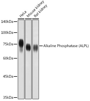
Western blot analysis of various lysates using Alkaline Phosphatase (ALPL) Rabbit pAb (CAB1080) at 1:1000 dilution. Secondary antibody: HRP-conjugated Goat anti-Rabbit IgG (H+L) (AS014) at 1:10000 dilution. Lysates/proteins: 25μg per lane. Blocking buffer: 3% nonfat dry milk in TBST. Detection: ECL Basic Kit (AbGn00020). Exposure time: 10s.
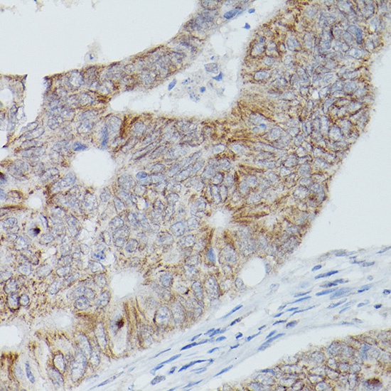
Immunohistochemistry analysis of paraffin-embedded Human colon carcinoma using Alkaline Phosphatase (ALPL) Rabbit pAb (CAB1080) at dilution of 1:50 (40x lens). High pressure antigen retrieval performed with 0.01M Citrate buffer (pH 6.0) prior to IHC staining.
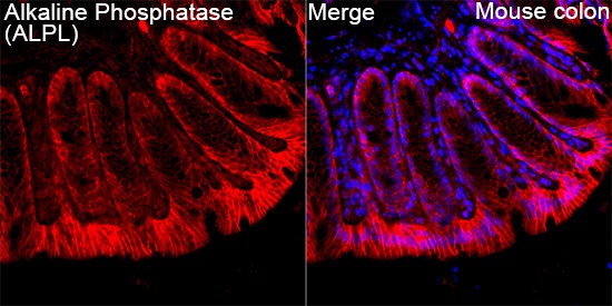
Immunofluorescence analysis of paraffin-embedded Mouse colon tissue using Alkaline Phosphatase (ALPL) Rabbit pAb (CAB1080) at a dilution of 1:200 (40x lens). Secondary antibody: Cy3 Goat Anti-Rabbit IgG (H+L)(AS007) at 1:500 dilution. Blue: DAPI for nuclear staining. Perform high pressure antigen retrieval with 0.01 M citrate buffer (pH 6.0) prior to IF staining.
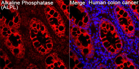
Immunofluorescence analysis of Human colon cancer tissue using Alkaline Phosphatase (ALPL) Rabbit pAb (CAB1080) at a dilution of 1:200 (40x lens). Secondary antibody: Cy3-conjugated Goat anti-Rabbit IgG (H+L)(AS007) at 1:500 dilution. Blue: DAPI for nuclear staining. High pressure antigen retrieval performed with 0.01M Citrate Buffer (pH 6.0) prior to IF staining.


")
")
