The AMPKbeta1 Monoclonal Antibody (CAB4344) is a high-quality antibody developed for reliable detection and analysis of target proteins. This antibody, produced in rabbits, specifically binds to the AMPKbeta1 subunit, allowing for precise detection and analysis in various cell types and tissues.The AMPK signaling pathway plays a vital role in cellular energy homeostasis, regulating processes such as metabolism, cell growth, and autophagy. Dysregulation of AMPK has been implicated in a variety of diseases, including diabetes, cancer, and neurological disorders.
This antibody is validated for use in WB, IHC-P, IF/ICC, ELISA applications and has demonstrated reactivity against Human, Mouse, Rat samples.
Product Name:
AMPKbeta1 Monoclonal Antibody
SKU:
CAB4344
Size:
20μL, 100μL
Reactivity:
Human, Mouse, Rat
Clone Number:
ARC1061
Conjugate:
Unconjugated
Immunogen:
Synthetic peptide. This information is considered to be commercially sensitive.
Recommended starting concentration is 1 μg/mL. Please optimize the concentration based on your specific assay requirements.
Synonyms:
AMPK, HAMPKb, AMPKβ1
Positive Sample:
Mouse brain, Rat lung, Rat kidney
Cellular Localization:
Cytoplasm, Cytosol, Nucleoplasm, Nucleus.
Calculated MW:
30kDa
Observed MW:
38kDa
The protein encoded by this gene is a regulatory subunit of the AMP-activated protein kinase (AMPK). AMPK is a heterotrimer consisting of an alpha catalytic subunit, and non-catalytic beta and gamma subunits. AMPK is an important energy-sensing enzyme that monitors cellular energy status. In response to cellular metabolic stresses, AMPK is activated, and thus phosphorylates and inactivates acetyl-CoA carboxylase (ACC) and beta-hydroxy beta-methylglutaryl-CoA reductase (HMGCR), key enzymes involved in regulating de novo biosynthesis of fatty acid and cholesterol. This subunit may be a positive regulator of AMPK activity. The myristoylation and phosphorylation of this subunit have been shown to affect the enzyme activity and cellular localization of AMPK. This subunit may also serve as an adaptor molecule mediating the association of the AMPK complex.
Purification Method
Affinity purification
Gene ID
5564
RRID
AB_2863240
Buffer Information
Store at -20℃. Avoid freeze / thaw cycles. Buffer: PBS containing 50% glycerol and 0.05% BSA, preserved with proclin300 or sodium azide, pH 7.3.
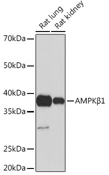
Western blot analysis of various lysates using AMPKβ1 Rabbit mAb (CAB4344) at 1:1000 dilution. Secondary antibody: HRP-conjugated Goat anti-Rabbit IgG (H+L) (CABS014) at 1:10000 dilution. Lysates/proteins: 25μg per lane. Blocking buffer: 3% nonfat dry milk in TBST. Detection: ECL Basic Kit (AbGn00020). Exposure time: 60s.
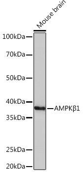
Western blot analysis of lysates from Mouse brain, using AMPKβ1 Rabbit mAb (CAB4344) at 1:1000 dilution. Secondary antibody: HRP-conjugated Goat anti-Rabbit IgG (H+L) (CABS014) at 1:10000 dilution. Lysates/proteins: 25μg per lane. Blocking buffer: 3% nonfat dry milk in TBST. Detection: ECL Basic Kit (AbGn00020). Exposure time: 3min.
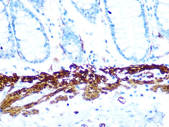
Immunohistochemistry analysis of paraffin-embedded Human colon using AMPKβ1 Rabbit mAb (CAB4344) at dilution of 1:100 (40x lens). Microwave antigen retrieval performed with 0.01M PBS Buffer (pH 7.2) prior to IHC staining.
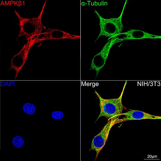
Confocal imaging of NIH/3T3 cells using AMPKβ1 Rabbit mAb (CAB4344,dilution 1:100)(Red). The cells were counterstained with α-Tubulin Mouse mAb (AC012,dilution 1:400) (Green). DAPI was used for nuclear staining (blue). Objective: 100x.