The AMT Antibody (CAB9926) is a high-quality antibody developed for reliable detection and analysis of target proteins. This antibody is derived from rabbits and shows high reactivity with human samples, making it suitable for a wide range of research applications.The AMT Polyclonal Antibody is specifically designed for use in Western blotting, enabling researchers to detect and analyze AMT protein levels in different cell types. By binding to the AMT protein, this antibody allows for precise and accurate measurements, making it an essential tool for studies in cell biology, molecular biology, and disease research.
This antibody is validated for use in WB, IF/ICC, ELISA applications and has demonstrated reactivity against Human, Mouse, Rat samples.
Product Name:
AMT Antibody
SKU:
CAB9926
Size:
20μL, 100μL
Reactivity:
Human, Mouse, Rat
Conjugate:
Unconjugated
Immunogen:
Recombinant protein (or fragment).This information is considered to be commercially sensitive.
Recommended starting concentration is 1 μg/mL. Please optimize the concentration based on your specific assay requirements.
Synonyms:
GCE, NKH, GCST, GCVT, AMT
Positive Sample:
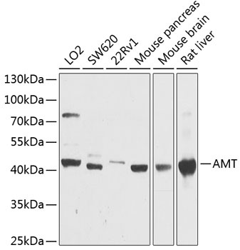
LO2, SW620, 22Rv1, Mouse pancreas, Mouse brain, Rat liver
Cellular Localization:
Mitochondrion.
Calculated MW:
44kDa
Observed MW:
44kDa
This gene encodes one of four critical components of the glycine cleavage system. Mutations in this gene have been associated with glycine encephalopathy. Multiple transcript variants encoding different isoforms have been found for this gene.
Purification Method
Affinity purification
Gene ID
275
RRID
AB_2768353
Buffer Information
Store at -20℃. Avoid freeze / thaw cycles. Buffer: PBS containing 50% glycerol, preserved with proclin300 or sodium azide, pH 7.3.
Western blot analysis of various lysates using AMT Rabbit pAb (CAB9926) at 1:1000 dilution. Secondary antibody: HRP-conjugated Goat anti-Rabbit IgG (H+L) (CABS014) at 1:10000 dilution. Lysates/proteins: 25μg per lane. Blocking buffer: 3% nonfat dry milk in TBST. Detection: ECL Basic Kit (AbGn00020). Exposure time: 90s.
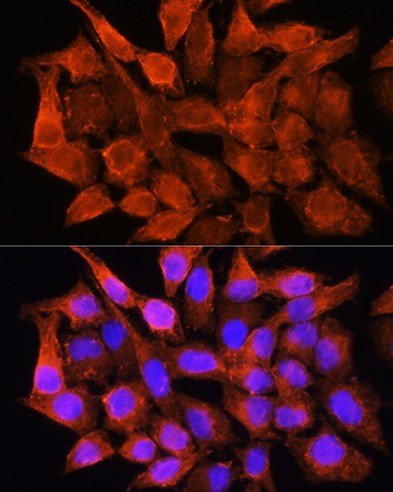
Immunofluorescence analysis of HeLa cells using AMT Rabbit pAb (CAB9926) at dilution of 1:100. Secondary antibody: Cy3-conjugated Goat anti-Rabbit IgG (H+L) (CABS007) at 1:500 dilution. Blue: DAPI for nuclear staining.