The Acetyl-Histone H3-K23 Monoclonal Antibody (CAB2771) is a high-quality antibody developed for reliable detection and analysis of target proteins. This antibody, produced using advanced technology, specifically targets acetylation at lysine 23 on histone H3, a key modification involved in chromatin structure and transcriptional regulation.Histone acetylation at lysine 23 has been linked to various biological processes, including DNA repair, cell cycle regulation, and development. This antibody is highly specific and validated for use in various applications, including immunofluorescence, chromatin immunoprecipitation (ChIP), and immunohistochemistry.
This antibody is validated for use in WB, IHC-P, IF/ICC, IP, ChIP, ELISA, DB applications and has demonstrated reactivity against Human, Mouse, Rat, Other (Wide Range Predicted) samples.
Product Name:
Acetyl-Histone H3-K23 Monoclonal Antibody
SKU:
CAB2771
Size:
20μL, 100μL
Reactivity:
Human, Mouse, Rat, Other (Wide Range Predicted)
Clone Number:
ARC53670
Conjugate:
Unconjugated
Immunogen:
Synthetic peptide. This information is considered to be commercially sensitive.
0.5μg-4μg antibody for 200μg-400μg extracts of whole cells
ELISA
Recommended starting concentration is 1 μg/mL. Please optimize the concentration based on your specific assay requirements.
ChIP
2μg antibody for 5μg-10μg of Chromatin
Positive Sample:
HeLa treated with TSA, NIH/3T3 treated with TSA, C2C12 treated with TSA,
Cellular Localization:
Chromosome, Nucleus.
Calculated MW:
15kDa
Observed MW:
17kDa
Core component of nucleosome. Nucleosomes wrap and compact DNA into chromatin, limiting DNA accessibility to the cellular machineries which require DNA as a template. Histones thereby play a central role in transcription regulation, DNA repair, DNA replication and chromosomal stability. DNA accessibility is regulated via a complex set of post-translational modifications of histones, also called histone code, and nucleosome remodeling.
Purification Method
Affinity purification
Gene ID
8290 8350
Buffer Information
Store at -20℃. Avoid freeze / thaw cycles. Buffer: PBS containing 50% glycerol and 0.05% BSA, preserved with proclin300 or sodium azide, pH 7.3.
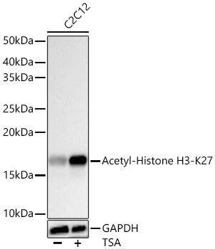
Western blot analysis of lysates from C2C12 cells using Acetyl-Histone H3-K27 Rabbit mAb (CAB2771) at 1:100000 dilution. C2C12 cells were treated with TSA (1 uM) at 37℃ for 18 hours. Secondary antibody: HRP-conjugated Goat anti-Rabbit IgG (H+L) (CABS014) at 1:10000 dilution. Lysates/proteins: 20 μg per lane. Blocking buffer: 3% nonfat dry milk in TBST. Detection: ECL Basic Kit (AbGn00020). Exposure time: 30s.
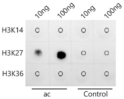
Dot-blot analysis of all sorts of peptides using Acetyl-Histone H3-K27 Rabbit mAb (CAB2771) at 1:200000 dilution.
Immunohistochemistry analysis of paraffin-embedded Human thyroid cancer tissue using Acetyl-Histone H3-K27 Rabbit mAb (CAB2771) at a dilution of 1:1000 (40x lens). High pressure antigen retrieval was performed with 0.01 M citrate buffer (pH 6.0) prior to IHC staining.
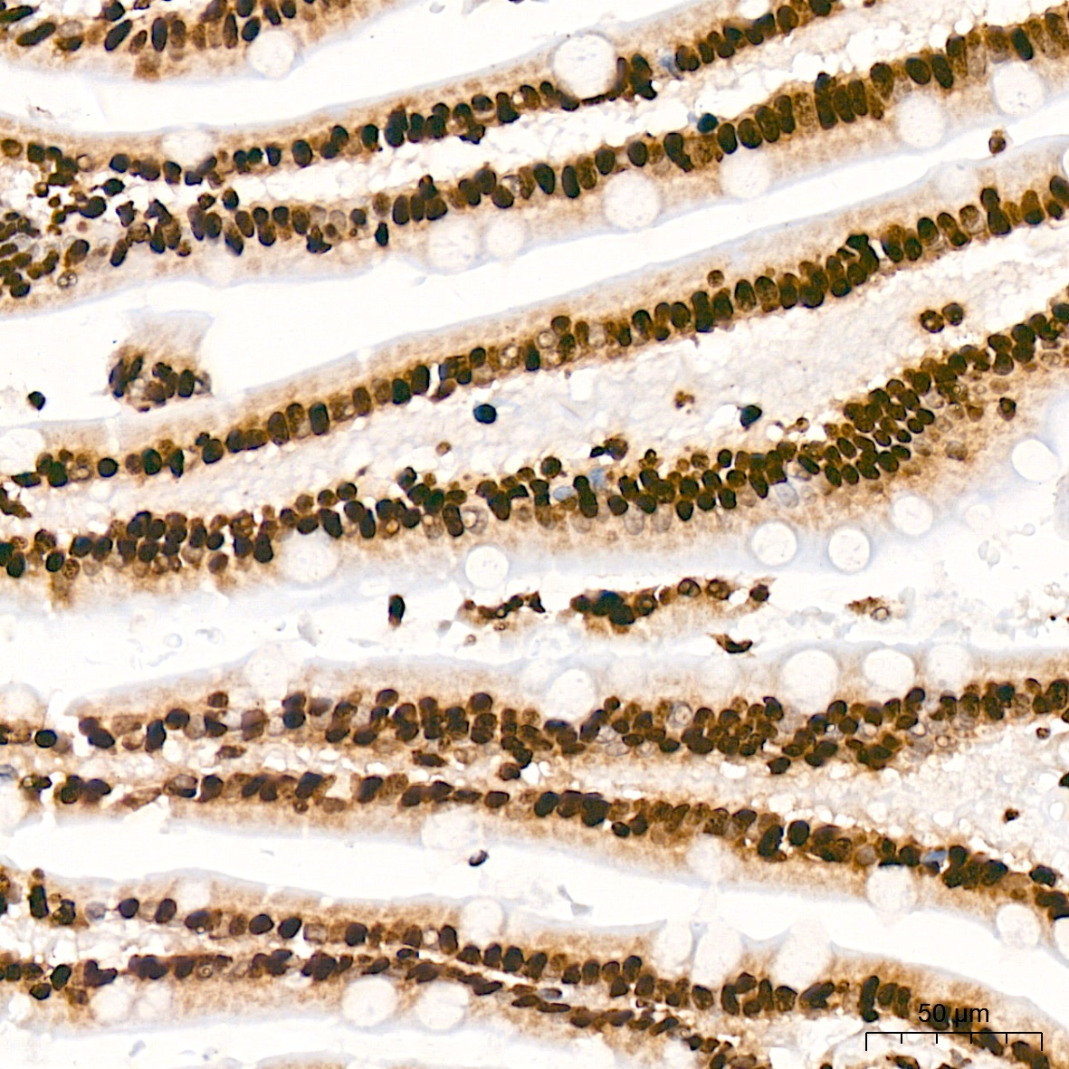
Immunohistochemistry analysis of paraffin-embedded Human small intestine tissue using Acetyl-Histone H3-K27 Rabbit mAb (CAB2771) at a dilution of 1:1000 (40x lens). High pressure antigen retrieval was performed with 0.01 M citrate buffer (pH 6.0) prior to IHC staining.
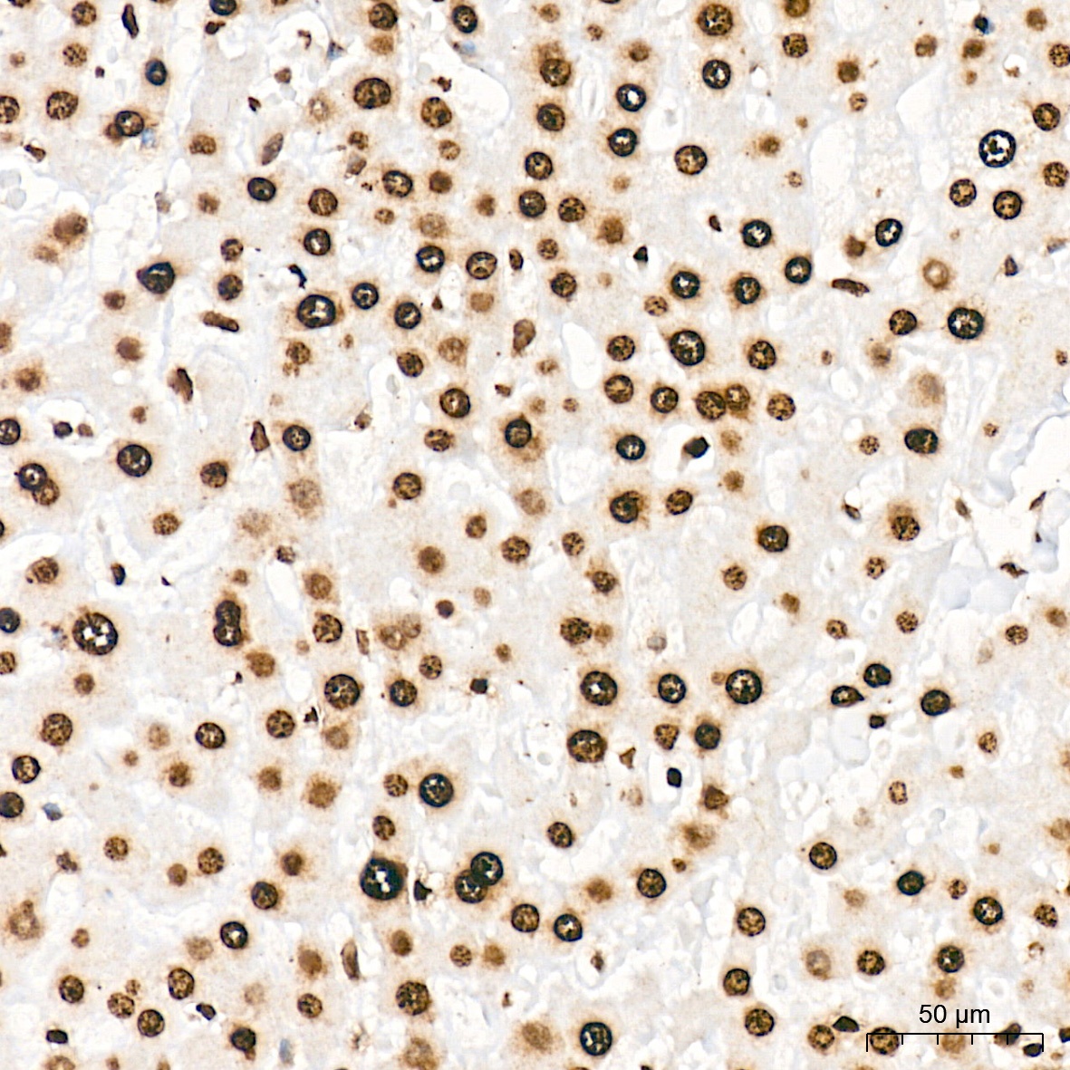
Immunohistochemistry analysis of paraffin-embedded Human liver tissue using Acetyl-Histone H3-K27 Rabbit mAb (CAB2771) at a dilution of 1:1000 (40x lens). High pressure antigen retrieval was performed with 0.01 M citrate buffer (pH 6.0) prior to IHC staining.
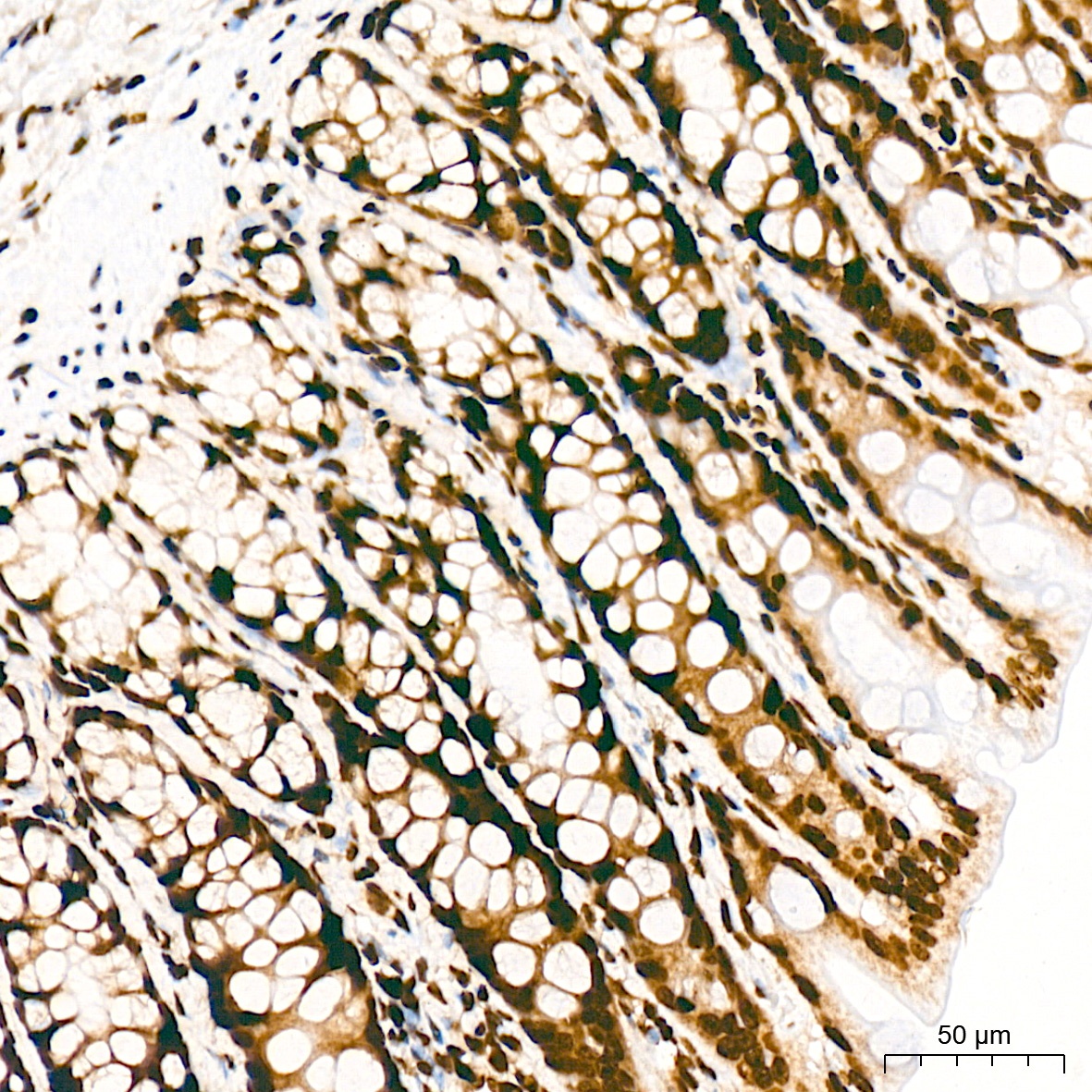
Immunohistochemistry analysis of paraffin-embedded Rat colon tissue using Acetyl-Histone H3-K27 Rabbit mAb (CAB2771) at a dilution of 1:1000 (40x lens). High pressure antigen retrieval was performed with 0.01 M citrate buffer (pH 6.0) prior to IHC staining.
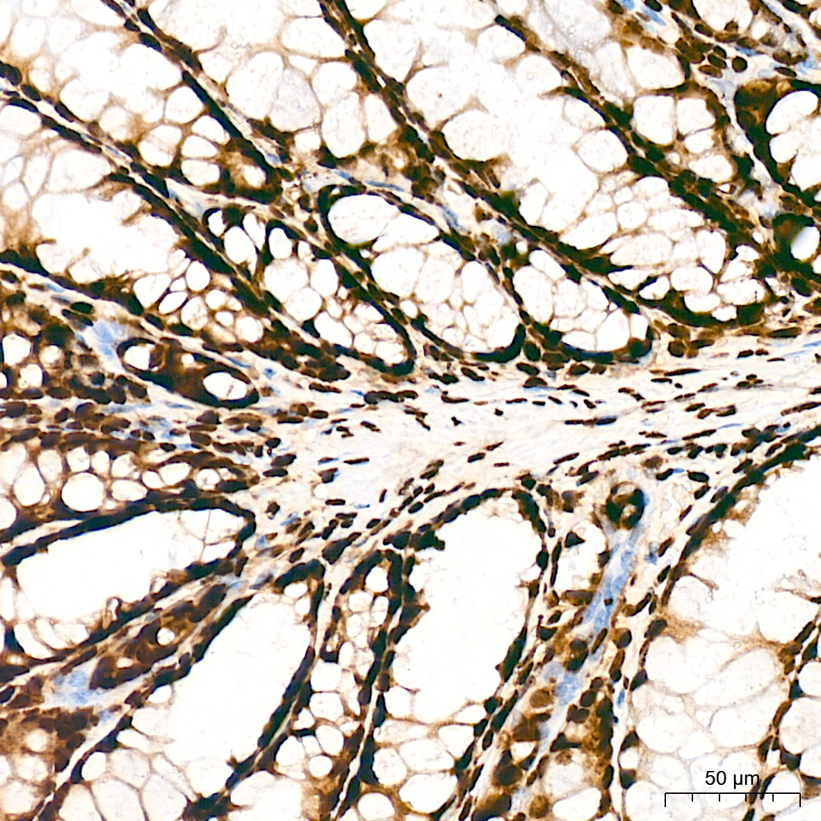
Immunohistochemistry analysis of paraffin-embedded Mouse colon tissue using Acetyl-Histone H3-K27 Rabbit mAb (CAB2771) at a dilution of 1:1000 (40x lens). High pressure antigen retrieval was performed with 0.01 M citrate buffer (pH 6.0) prior to IHC staining.
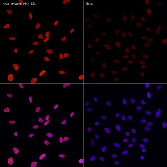
Immunofluorescence analysis of HeLa treated with TSA and HeLa cells using Acetyl-Histone H3-K27 Rabbit mAb (CAB2771) at dilution of 1:50 (40x lens). Secondary antibody: Cy3-conjugated Goat anti-Rabbit IgG (H+L) (CABS007) at 1:500 dilution. Blue: DAPI for nuclear staining.
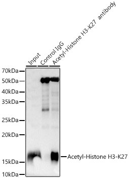
Immunoprecipitation analysis of 600 μg extracts of HeLa cells treated with TSA using 5 μg Acetyl-Histone H3-K27 Rabbit mAb(CAB2771). Western blot was performed from the immunoprecipitate using Acetyl-Histone H3-K27 antibody (CAB2771) at a dilution of 1:50000.
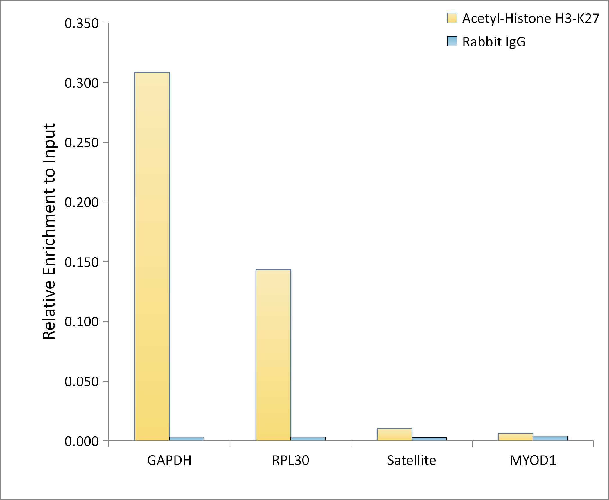
Chromatin immunoprecipitation analysis of extracts of HeLa cells, using Acetyl-Histone H3-K27 antibody (CAB2771) and rabbit IgG.The amount of immunoprecipitated DNA was checked by quantitative PCR. Histogram was constructed by the ratios of the immunoprecipitated DNA to the input.