The ACVR1B Monoclonal Antibody (CAB2279) is a high-quality antibody developed for reliable detection and analysis of target proteins. This antibody, produced in mice, exhibits high specificity towards human samples and is suitable for use in applications such as immunofluorescence and flow cytometry.ACVR1B plays a vital role in signal transduction pathways related to cell proliferation and differentiation, making it a key player in development and disease.
This antibody is validated for use in WB, IHC-P, IF/ICC, ELISA applications and has demonstrated reactivity against Human, Mouse, Rat samples.
Product Name:
ACVR1B Monoclonal Antibody
SKU:
CAB2279
Size:
20μL, 100μL
Reactivity:
Human, Mouse, Rat
Clone Number:
ARC1899
Conjugate:
Unconjugated
Immunogen:
Synthetic peptide. This information is considered to be commercially sensitive.
Recommended starting concentration is 1 μg/mL. Please optimize the concentration based on your specific assay requirements.
Synonyms:
ALK4, SKR2, ACTRIB, ACVRLK4, ACVR1B
Positive Sample:
293T, HepG2, U-87MG, Mouse brain, Rat brain
Cellular Localization:
Cell Surface, Cytosol, Plasma Membrane.
Calculated MW:
57kDa
Observed MW:
57kDa
This gene encodes an activin A type IB receptor. Activins are dimeric growth and differentiation factors which belong to the transforming growth factor-beta (TGF-beta) superfamily of structurally related signaling proteins. Activins signal through a heteromeric complex of receptor serine kinases which include at least two type I and two type II receptors. This protein is a type I receptor which is essential for signaling. Mutations in this gene are associated with pituitary tumors. Alternate splicing results in multiple transcript variants.
Purification Method
Affinity purification
Gene ID
91
RRID
AB_2862987
Buffer Information
Store at -20℃. Avoid freeze / thaw cycles. Buffer: PBS containing 50% glycerol and 0.05% BSA, preserved with proclin300 or sodium azide, pH 7.3.
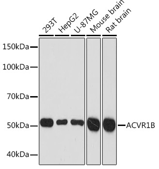
Western blot analysis of various lysates using ACVR1B Rabbit mAb (CAB2279) at 1:1000 dilution. Secondary antibody: HRP-conjugated Goat anti-Rabbit IgG (H+L) (CABS014) at 1:10000 dilution. Lysates/proteins: 25μg per lane. Blocking buffer: 3% nonfat dry milk in TBST. Detection: ECL Basic Kit (AbGn00020). Exposure time: 3min.
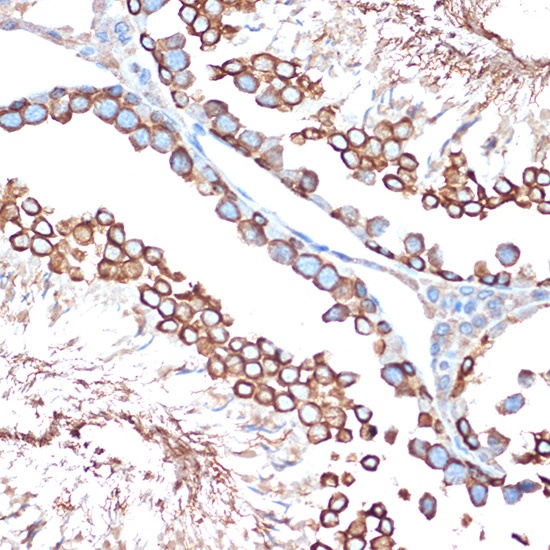
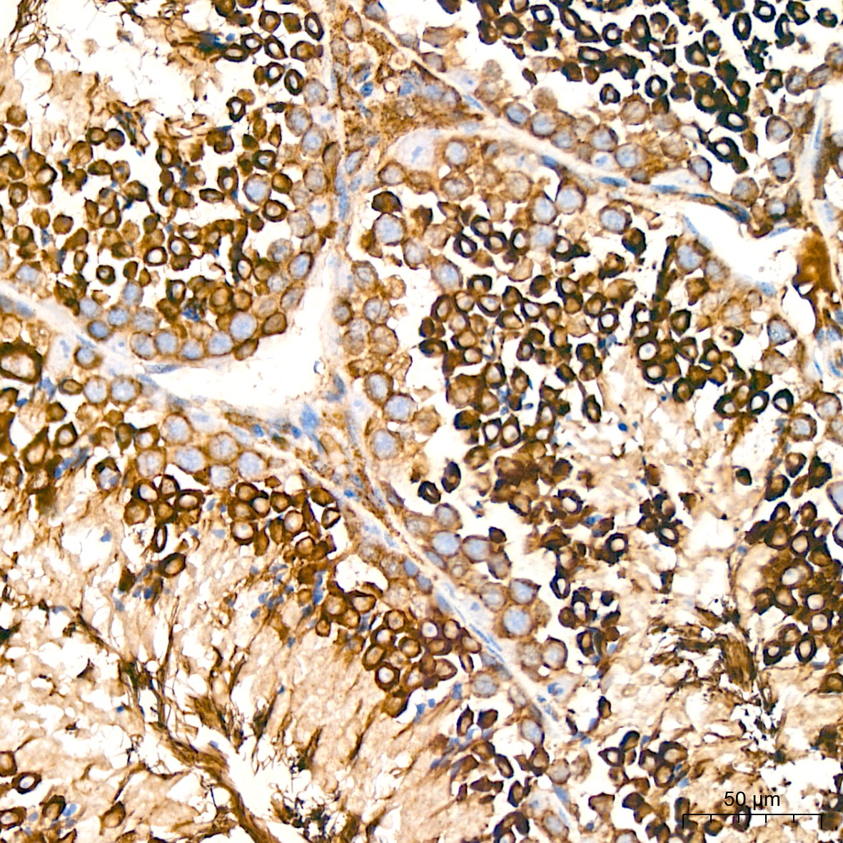
Immunohistochemistry analysis of paraffin-embedded Rat testis using ACVR1B Rabbit mAb (CAB2279) at dilution of 1:100 (40x lens). Microwave antigen retrieval performed with 0.01M Tris/EDTA Buffer (pH 9.0) prior to IHC staining.
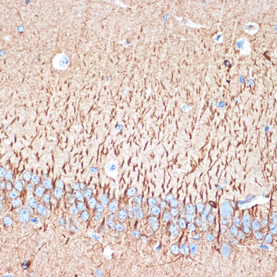
Immunohistochemistry analysis of paraffin-embedded Mouse brain using ACVR1B Rabbit mAb (CAB2279) at dilution of 1:100 (40x lens). Microwave antigen retrieval performed with 0.01M Tris/EDTA Buffer (pH 9.0) prior to IHC staining.
Immunohistochemistry analysis of paraffin-embedded Human cervix cancer tissue using ACVR1B Rabbit mAb (CAB22347) at a dilution of 1:200 (40x lens). High pressure antigen retrieval performed with 0.01M Citrate buffer (pH 6.0) prior to IHC staining.
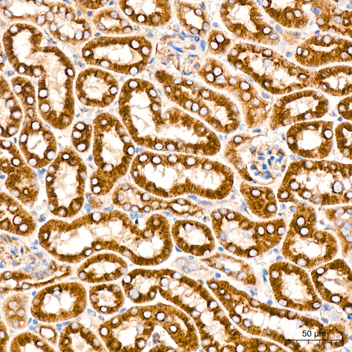
Immunohistochemistry analysis of paraffin-embedded Mouse kidney tissue using ACVR1B Rabbit mAb (CAB22347) at a dilution of 1:200 (40x lens). High pressure antigen retrieval performed with 0.01M Citrate buffer (pH 6.0) prior to IHC staining.
Immunohistochemistry analysis of paraffin-embedded Mouse testis tissue using ACVR1B Rabbit mAb (CAB22347) at a dilution of 1:200 (40x lens). High pressure antigen retrieval performed with 0.01M Citrate buffer (pH 6.0) prior to IHC staining.
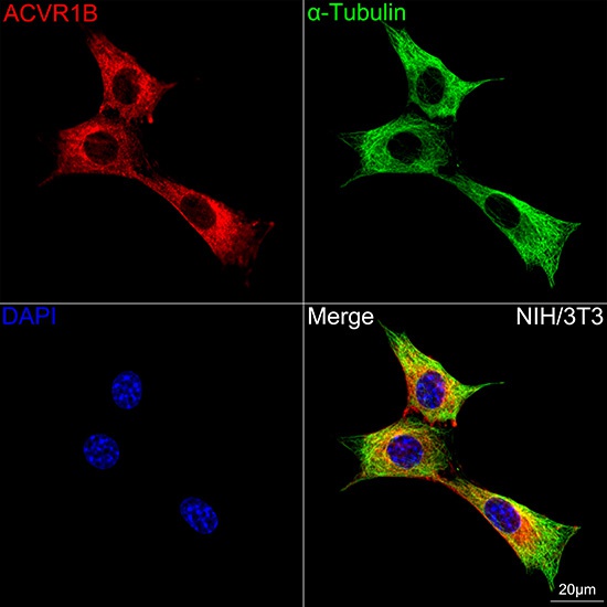
Confocal imaging of NIH/3T3 cells using ACVR1B Rabbit mAb (CAB2279,dilution 1:100) followed by a further incubation with Cy3 Goat Anti-Rabbit IgG (H+L) (CABS007,dilution 1:500)(Red).The cells were counterstained with α-Tubulin Mouse mAb (AC012, dilution 1:400) followed by incubation with ABflo® 488-conjugated Goat Anti-Mouse IgG (H+L) Ab (CABS076, dilution 1:500) (Green).DAPI was used for nuclear staining (Blue). Objective: 100x.