The ACVRL1 Monoclonal Antibody (CAB9755) is a high-quality antibody developed for reliable detection and analysis of target proteins. This gene encodes a type I cell-surface receptor for the TGF-beta superfamily of ligands. It shares with other type I receptors a high degree of similarity in serine-threonine kinase subdomains, a glycine- and serine-rich region (called the GS domain) preceding the kinase domain, and a short C-terminal tail. The encoded protein, sometimes termed ALK1, shares similar domain structures with other closely related ALK or activin receptor-like kinase proteins that form a subfamily of receptor serine/threonine kinases. Mutations in this gene are associated with hemorrhagic telangiectasia type 2, also known as Rendu-Osler-Weber syndrome 2.
This antibody is validated for use in WB, IF/ICC, ELISA applications and has demonstrated reactivity against Human, Mouse, Rat samples.
Product Name:
ACVRL1 Monoclonal Antibody
SKU:
CAB9755
Size:
100μL, 20μL
Reactivity:
Human, Mouse, Rat
Clone Number:
ARC1735
Conjugate:
Unconjugated
Immunogen:
Synthetic peptide. This information is considered to be commercially sensitive.
Tested Applications:
WBIF/ICCELISA
Recommended Dilution:
WB
1:1000 - 1:6000
IF/ICC
1:50 - 1:200
ELISA
Recommended starting concentration is 1 μg/mL. Please optimize the concentration based on your specific assay requirements.
This gene encodes a type I cell-surface receptor for the TGF-beta superfamily of ligands. It shares with other type I receptors a high degree of similarity in serine-threonine kinase subdomains, a glycine- and serine-rich region (called the GS domain) preceding the kinase domain, and a short C-terminal tail. The encoded protein, sometimes termed ALK1, shares similar domain structures with other closely related ALK or activin receptor-like kinase proteins that form a subfamily of receptor serine/threonine kinases. Mutations in this gene are associated with hemorrhagic telangiectasia type 2, also known as Rendu-Osler-Weber syndrome 2.
Purification Method
Affinity purification
Gene ID
94
RRID
AB_2863773
Buffer Information
Store at -20℃. Avoid freeze / thaw cycles. Buffer: PBS containing 50% glycerol and 0.05% BSA, preserved with proclin300 or sodium azide, pH 7.3.
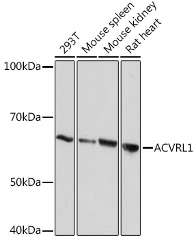
Western blot analysis of various lysates using ACVRL1 Rabbit mAb (CAB9755) at 1:1000 dilution. Secondary antibody: HRP-conjugated Goat anti-Rabbit IgG (H+L) (AS014) at 1:10000 dilution. Lysates/proteins: 25μg per lane. Blocking buffer: 3% nonfat dry milk in TBST. Detection: ECL Basic Kit (AbGn00020). Exposure time: 10s.
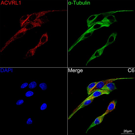
Confocal imaging of C6 cells using ACVRL1 Rabbit mAb (CAB9755, dilution 1:100) followed by a further incubation with Cy3 Goat Anti-Rabbit IgG (H+L) (AS007, dilution 1:500) (Red). The cells were counterstained with α-Tubulin Mouse mAb (AC012, dilution 1:400) followed by incubation with ABflo® 488-conjugated Goat Anti-Mouse IgG (H+L) Ab (AS076, dilution 1:500) (Green). DAPI was used for nuclear staining (Blue). Objective: 100x.