The AIF1/IBA1 Monoclonal Antibody (CAB19776) is a high-quality antibody developed for reliable detection and analysis of target proteins. This antibody, produced in rabbits, is highly specific to human samples and has been validated for use in Western blot applications. It binds specifically to the AIF1/IBA1 protein, allowing for detection and analysis in various cell types, making it ideal for studies in neuroscience, neuroinflammation, and neurodegenerative diseases.AIF1/IBA1 is known for its role in the activation and regulation of microglia and macrophages in response to injury or inflammation in the central nervous system.
This antibody is validated for use in WB, IHC-P, ELISA, IF-F, IF-P applications and has demonstrated reactivity against Human, Mouse, Rat samples.
Product Name:
AIF1/IBA1 Monoclonal Antibody
SKU:
CAB19776
Size:
20μL, 100μL
Reactivity:
Human, Mouse, Rat
Clone Number:
ARC2301
Conjugate:
Unconjugated
Immunogen:
Synthetic peptide. This information is considered to be commercially sensitive.
Recommended starting concentration is 1 μg/mL. Please optimize the concentration based on your specific assay requirements.
Synonyms:
G1, Iba1, AIF-1, D17H6S50E, AIF1/IBA1
Positive Sample:
THP-1, RAW 264.7, Mouse testis, Mouse spleen, Mouse brain, Rat lung, Rat testis, Rat spleen
Calculated MW:
17kDa
Observed MW:
17kDa
Enables actin filament binding activity and calcium ion binding activity. Involved in several processes, including Rac protein signal transduction; actin filament organization; and ruffle assembly. Acts upstream of or within actin filament bundle assembly. Located in several cellular components, including actin filament; phagocytic cup; and ruffle membrane. Is expressed in adrenal cortex; central nervous system; embryo mesenchyme; and retina. Human ortholog(s) of this gene implicated in type 1 diabetes mellitus. Orthologous to human AIF1 (allograft inflammatory factor 1).
Purification Method
Affinity purification
Gene ID
11629
RRID
AB_3711469
Buffer Information
Store at -20℃. Avoid freeze / thaw cycles. Buffer: PBS with 0.09% sodium azide,0.05% BSA,50% glycerol,pH7.3.
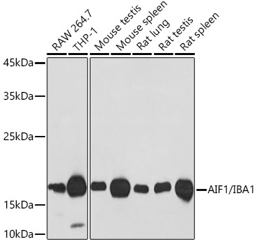
Western blot analysis of various lysates using AIF1/IBA1 Rabbit mAb (CAB19776) at 1:1000 dilution. Secondary antibody: HRP-conjugated Goat anti-Rabbit IgG (H+L) (CABS014) at 1:10000 dilution. Lysates/proteins: 25μg per lane. Blocking buffer: 3% nonfat dry milk in TBST. Detection: ECL Basic Kit (AbGn00020). Exposure time: 30s.
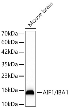
Western blot analysis of lysates from Mouse brain using AIF1/IBA1 Rabbit mAb (CAB19776) at 1:1000 dilution incubated overnight at 4℃. Secondary antibody: HRP-conjugated Goat anti-Rabbit IgG (H+L) (CABS014) at 1:10000 dilution. Lysates/proteins: 25 μg per lane. Blocking buffer: 3% nonfat dry milk in TBST. Detection: ECL Basic Kit (AbGn00020). Exposure time: 45s.
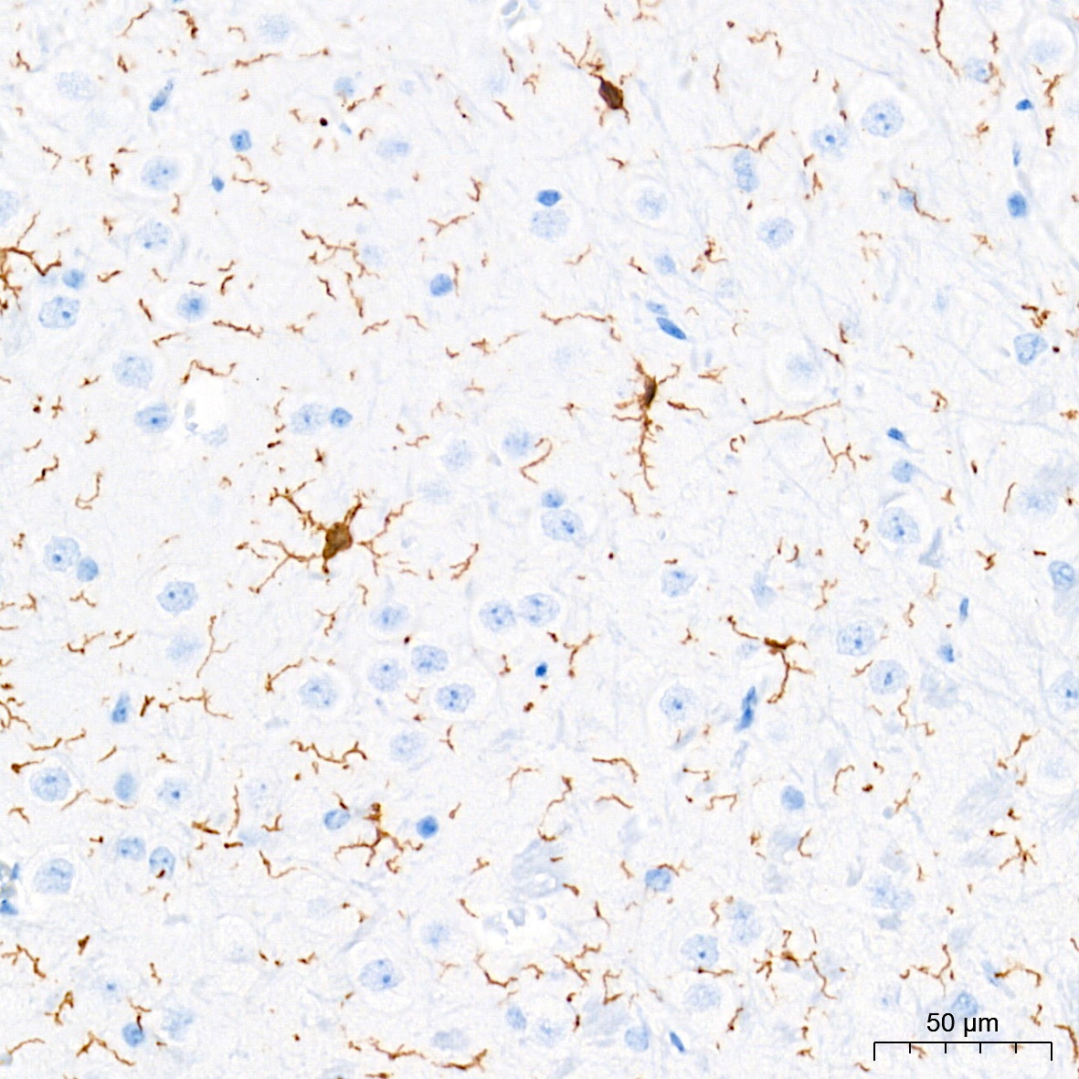
Immunohistochemistry analysis of paraffin-embedded Mouse brain tissue using AIF1/IBA1 Rabbit mAb (CAB19776) at a dilution of 1:20000 (40x lens). High pressure antigen retrieval performed with 0.01M Tris-EDTA Buffer (pH 9.0) prior to IHC staining.
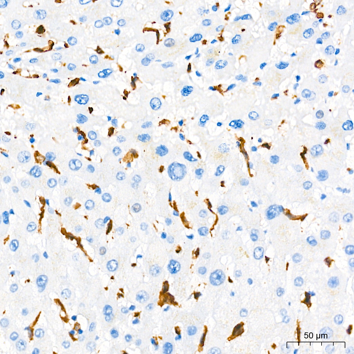
Immunohistochemistry analysis of paraffin-embedded Human liver tissue using AIF1/IBA1 Rabbit mAb (CAB19776) at a dilution of 1:20000 (40x lens). High pressure antigen retrieval performed with 0.01M Tris-EDTA Buffer (pH 9.0) prior to IHC staining.
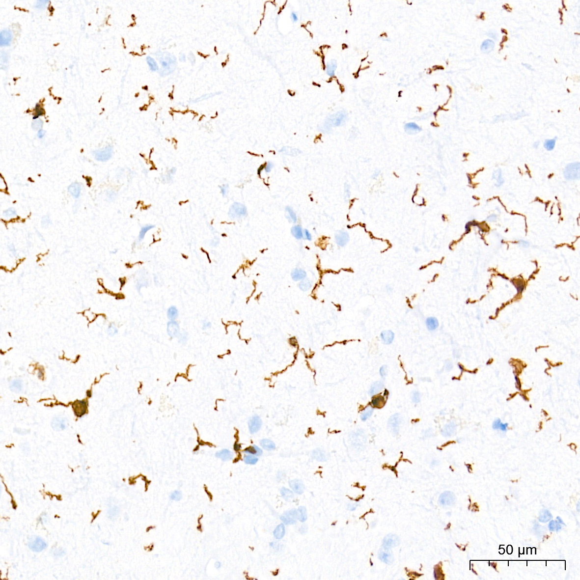
Immunohistochemistry analysis of paraffin-embedded Rat brain tissue using AIF1/IBA1 Rabbit mAb (CAB19776) at a dilution of 1:20000 (40x lens). High pressure antigen retrieval performed with 0.01M Tris-EDTA Buffer (pH 9.0) prior to IHC staining.
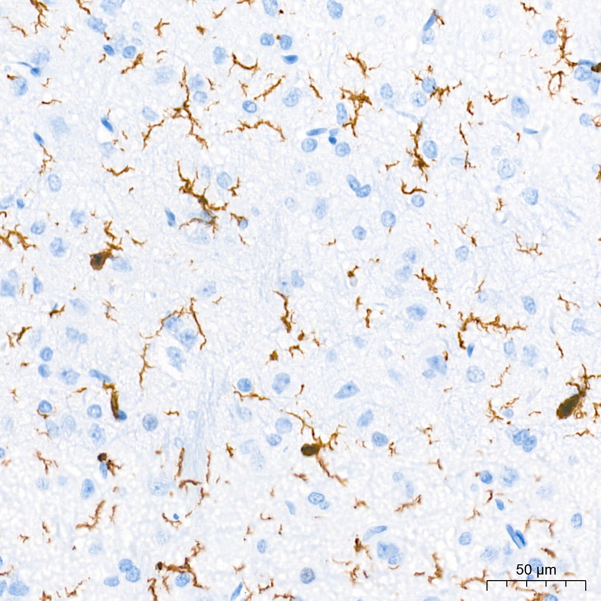
Immunohistochemistry analysis of paraffin-embedded Human brain tissue using AIF1/IBA1 Rabbit mAb (CAB19776) at a dilution of 1:20000 (40x lens). High pressure antigen retrieval performed with 0.01M Tris-EDTA Buffer (pH 9.0) prior to IHC staining.
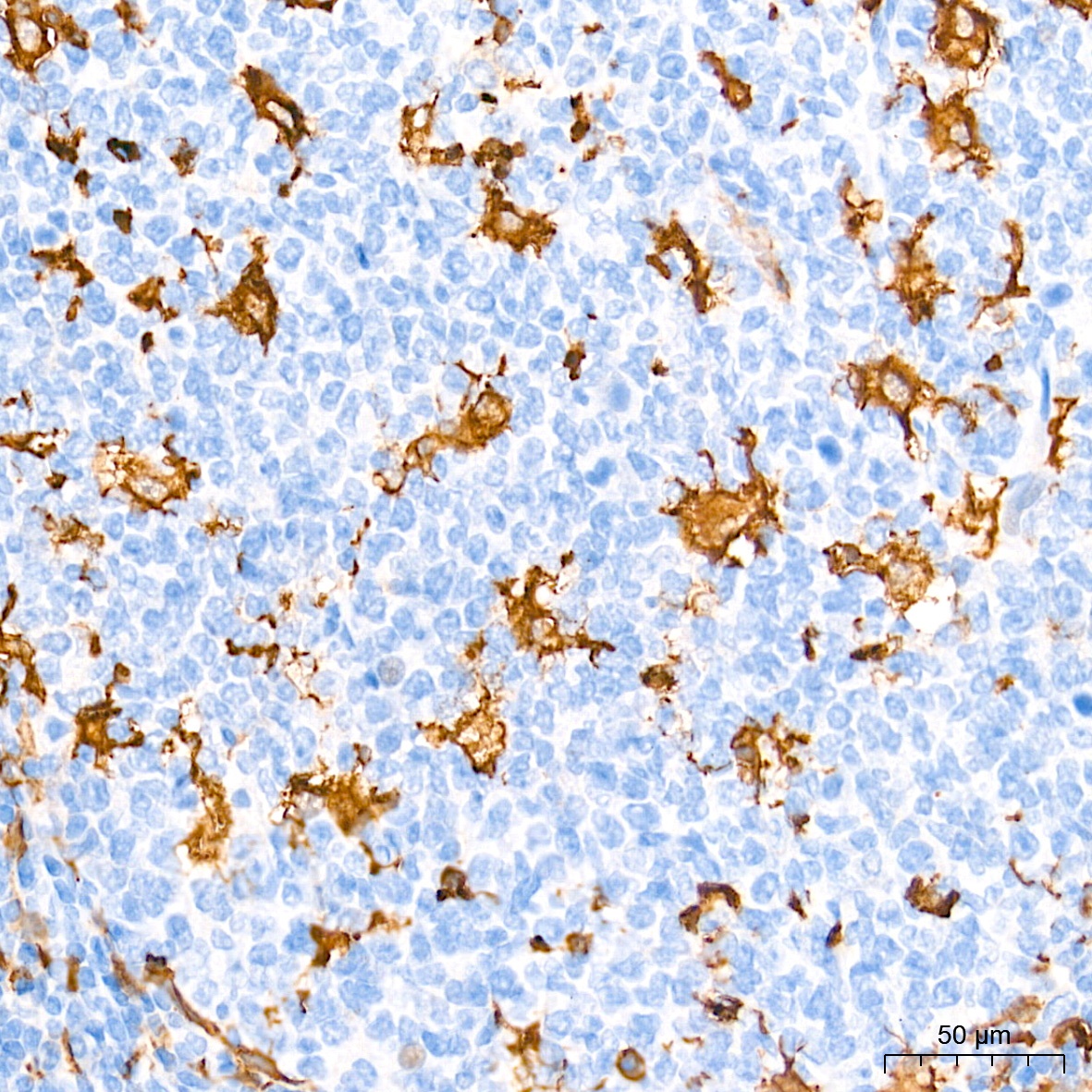
Immunohistochemistry analysis of paraffin-embedded Human tonsil tissue using AIF1/IBA1 Rabbit mAb (CAB19776) at a dilution of 1:20000 (40x lens). High pressure antigen retrieval performed with 0.01M Tris-EDTA Buffer (pH 9.0) prior to IHC staining.
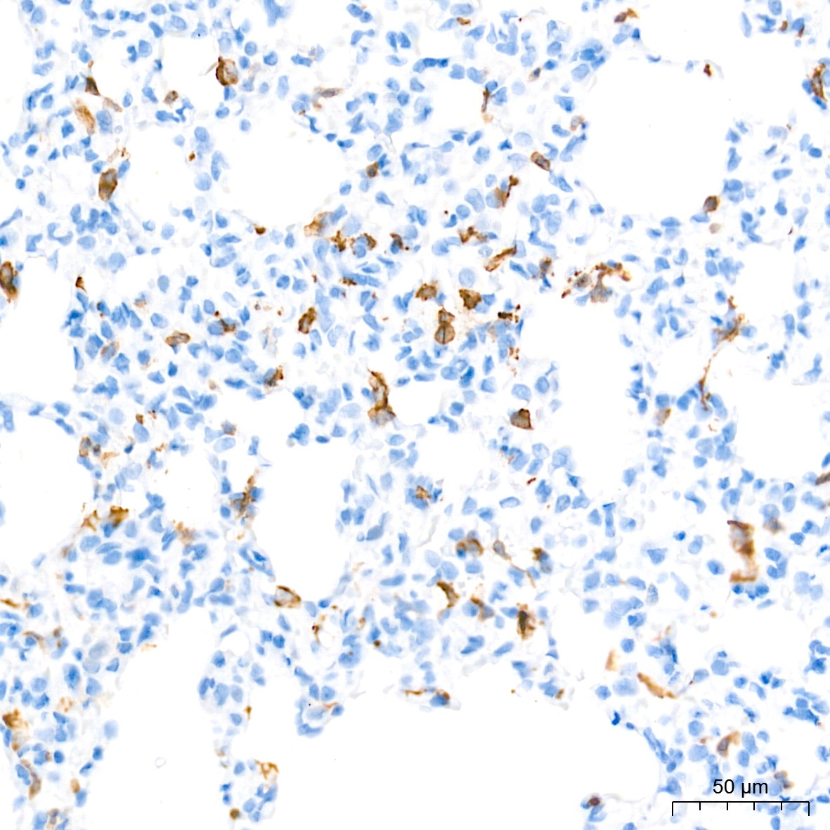
Immunohistochemistry analysis of paraffin-embedded Rat lung tissue using AIF1/IBA1 Rabbit mAb (CAB19776) at a dilution of 1:20000 (40x lens). High pressure antigen retrieval performed with 0.01M Tris-EDTA Buffer (pH 9.0) prior to IHC staining.
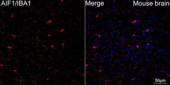
Confocal imaging of paraffin-embedded Mouse brain using AIF1/IBA1 Rabbit mAb (CAB19776, dilution 1:200) followed by a further incubation with Cy3 Goat Anti-Rabbit IgG (H+L) (CABS007, dilution 1:500) (Red). DAPI was used for nuclear staining (Blue). Objective: 40x.Perform microwave antigen retrieval with 0.01M citrate buffer (pH 6.0) prior to IF staining.
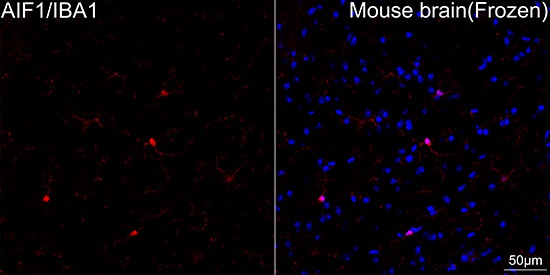
Confocal imaging of frozen sections Mouse brain tissue using AIF1/IBA1 Rabbit mAb (CAB19776, dilution 1:200) followed by a further incubation with Cy3 Goat Anti-Rabbit IgG (H+L) (CABS007, dilution 1:500) (Red). DAPI was used for nuclear staining (Blue). Microwave antigen retrieval performed with 0.01M Citrate Buffer (pH 6.0) prior to IF staining. Objective: 40x.
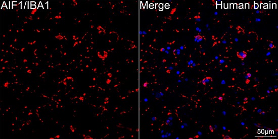
Confocal imaging of paraffin-embedded Human brain tissue using AIF1/IBA1 Rabbit mAb (CAB19776, dilution 1:200) followed by a further incubation with Cy3 Goat Anti-Rabbit IgG (H+L) (CABS007, dilution 1:500) (Red). DAPI was used for nuclear staining (Blue). High pressure antigen retrieval performed with 0.01M Citrate Buffer (pH 6.0) prior to IF staining. Objective: 40x.