The ARF5 Monoclonal Antibody (CAB3382) is a high-quality antibody developed for reliable detection and analysis of target proteins. This antibody, generated in rabbits, has been validated for use in applications like immunocytochemistry and immunofluorescence to detect and study ARF5 expression in different cell types.ARF5, a member of the ARF protein family, plays a crucial role in regulating vesicle formation and transport within cells, making it an important target for studying cellular trafficking pathways. Dysregulation of ARF5 function has been implicated in diseases such as cancer, neurodegenerative disorders, and infectious diseases.
This antibody is validated for use in WB, IF/ICC, ELISA applications and has demonstrated reactivity against Human, Mouse, Rat samples.
Product Name:
ARF5 Monoclonal Antibody
SKU:
CAB3382
Size:
20μL, 100μL
Reactivity:
Human, Mouse, Rat
Clone Number:
ARC1960
Conjugate:
Unconjugated
Immunogen:
Synthetic peptide. This information is considered to be commercially sensitive.
This gene is a member of the human ADP-ribosylation factor (ARF) gene family. These genes encode small guanine nucleotide-binding proteins that stimulate the ADP-ribosyltransferase activity of cholera toxin and play a role in vesicular trafficking and as activators of phospholipase D. The gene products include 6 ARF proteins and 11 ARF-like proteins and constitute 1 family of the RAS superfamily. The ARF proteins are categorized as class I (ARF1, ARF2,and ARF3), class II (ARF4 and ARF5) and class III (ARF6). The members of each class share a common gene organization.
Purification Method
Affinity purification
Gene ID
381
RRID
AB_2863047
Buffer Information
Store at -20℃. Avoid freeze / thaw cycles. Buffer: PBS containing 50% glycerol and 0.05% BSA, preserved with proclin300 or sodium azide, pH 7.3.
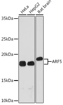
Western blot analysis of various lysates using ARF5 Rabbit mAb (CAB3382) at 1:1000 dilution. Secondary antibody: HRP-conjugated Goat anti-Rabbit IgG (H+L) (CABS014) at 1:10000 dilution. Lysates/proteins: 25μg per lane. Blocking buffer: 3% nonfat dry milk in TBST. Detection: ECL Basic Kit (AbGn00020). Exposure time: 3min.
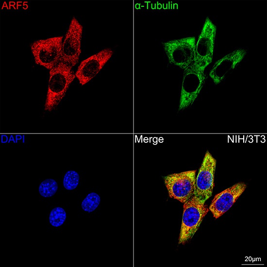
Confocal imaging of NIH/3T3 cells using ARF5 Rabbit mAb (CAB3382, dilution 1:100) followed by a further incubation with Cy3 Goat Anti-Rabbit IgG (H+L) (CABS007, dilution 1:500) (Red). The cells were counterstained with α-Tubulin Mouse mAb (AC012, dilution 1:400) followed by incubation with ABflo® 488-conjugated Goat Anti-Mouse IgG (H+L) Ab (CABS076, dilution 1:500) (Green). DAPI was used for nuclear staining (Blue). Objective: 100x.