The CD274 Antibody (CAB20481) is a high-quality antibody developed for reliable detection and analysis of target proteins. The protein encoded by this gene is an immune inhibitory receptor ligand that is expressed by hematopoietic and non-hematopoietic cells, such as T cells and B cells and various types of tumor cells. The encoded protein is a type I transmembrane protein that has immunoglobulin V-like and C-like domains. Interaction of this ligand with its receptor inhibits T-cell activation and cytokine production. During infection or inflammation of normal tissue, this interaction is important for preventing autoimmunity by maintaining homeostasis of the immune response. In tumor microenvironments, this interaction provides an immune escape for tumor cells through cytotoxic T-cell inactivation. Mice deficient for this gene display a variety of phenotypes including decreased allogeneic fetal survival rates and severe experimental autoimmune encephalomyelitis. RRID Gene ID 60533 Swiss Prot Synonym B7h1; Pdl1; Pdcd1l1; Pdcd1lg1; A530045L16Rik; 74
This antibody is validated for use in WB, ELISA applications and has demonstrated reactivity against Human, Mouse, Rat samples.
Product Name:
CD274 Antibody
SKU:
CAB20481
Size:
100μL, 20μL
Reactivity:
Human, Mouse, Rat
Clone Number:
-
Conjugate:
Unconjugated
Immunogen:
Recombinant protein (or fragment).This information is considered to be commercially sensitive.
Tested Applications:
WBELISA
Recommended Dilution:
WB
1:500 - 1:1000
ELISA
Recommended starting concentration is 1 μg/mL. Please optimize the concentration based on your specific assay requirements.
Synonyms:
B7h1, Pdl1, Pdcd1l1, Pdcd1lg1, A530045L16Rik, 74
Positive Sample:
A-549, Mouse skeletal muscle, Rat skeletal muscle
Cellular Localization:
Actin Cytoskeleton, Cell Surface, External Side Of Plasma Membrane, Extracellular Exosome, Nucleoplasm, Plasma Membrane.
Calculated MW:
33kDa
Observed MW:
40-50kDa
The protein encoded by this gene is an immune inhibitory receptor ligand that is expressed by hematopoietic and non-hematopoietic cells, such as T cells and B cells and various types of tumor cells. The encoded protein is a type I transmembrane protein that has immunoglobulin V-like and C-like domains. Interaction of this ligand with its receptor inhibits T-cell activation and cytokine production. During infection or inflammation of normal tissue, this interaction is important for preventing autoimmunity by maintaining homeostasis of the immune response. In tumor microenvironments, this interaction provides an immune escape for tumor cells through cytotoxic T-cell inactivation. Mice deficient for this gene display a variety of phenotypes including decreased allogeneic fetal survival rates and severe experimental autoimmune encephalomyelitis. RRID Gene ID 60533 Swiss Prot Synonym B7h1; Pdl1; Pdcd1l1; Pdcd1lg1; A530045L16Rik; 74
Purification Method:
Affinity purification
Gene ID:
60533
RRID:
-
Buffer Information:
Store at -20℃. Avoid freeze / thaw cycles. Buffer: PBS containing 50% glycerol, preserved with proclin300 or sodium azide, pH 7.3.
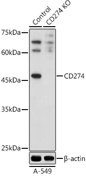
Western blot analysis of lysates from wild type (WT) and CD274 knockout (KO) A-549 cells, using [KO Validated] CD274 Rabbit pAb (CAB20481) at 1:500 dilution. Secondary antibody: HRP-conjugated Goat anti-Rabbit IgG (H+L) (AS014) at 1:10000 dilution. Lysates/proteins: 25μg per lane. Blocking buffer: 3% nonfat dry milk in TBST. Detection: ECL Basic Kit (AbGn00020). Exposure time: 180s.
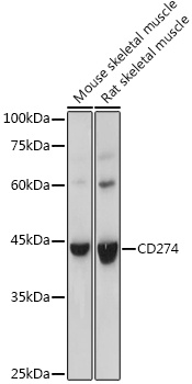
Western blot analysis of various lysates using [KO Validated] CD274 Rabbit pAb (CAB20481) at 1:500 dilution. Secondary antibody: HRP-conjugated Goat anti-Rabbit IgG (H+L) (AS014) at 1:10000 dilution. Lysates/proteins: 25μg per lane. Blocking buffer: 3% nonfat dry milk in TBST. Detection: ECL Basic Kit (AbGn00020). Exposure time: 30s.