The CD79B Monoclonal Antibody (CAB4362) is a high-quality antibody developed for reliable detection and analysis of target proteins. This antibody, generated in rabbits, has high specificity for human samples and is validated for use in various applications, including Western blot and immunohistochemistry. By binding to the CD79B protein, this antibody enables researchers to detect and analyze CD79B expression in different cell types, making it ideal for studies in immunology and cancer research.CD79B, also known as B-cell antigen receptor complex-associated protein beta chain, is essential for B-cell development and activation.
This antibody is validated for use in WB, IHC-P, ELISA, IF-P applications and has demonstrated reactivity against Human, Mouse, Rat samples.
Product Name:
CD79B Monoclonal Antibody
SKU:
CAB4362
Size:
20μL, 100μL
Reactivity:
Human, Mouse, Rat
Clone Number:
ARC52688
Conjugate:
Unconjugated
Immunogen:
Synthetic peptide. This information is considered to be commercially sensitive.
Recommended starting concentration is 1 μg/mL. Please optimize the concentration based on your specific assay requirements.
Synonyms:
B29, IGB, AGM6, Igbeta, CD79B
Positive Sample:
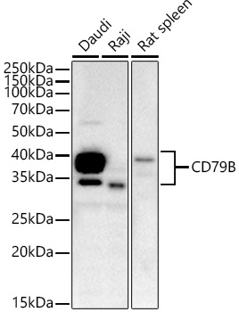
Daudi, Raji, Rat spleen
Cellular Localization:
External Side Of Plasma Membrane, Extracellular Exosome, Plasma Membrane.
Calculated MW:
26kDa
Observed MW:
30-50kDa
The B lymphocyte antigen receptor is a multimeric complex that includes the antigen-specific component, surface immunoglobulin (Ig). Surface Ig non-covalently associates with two other proteins, Ig-alpha and Ig-beta, which are necessary for expression and function of the B-cell antigen receptor. This gene encodes the Ig-beta protein of the B-cell antigen component. Alternatively spliced transcript variants encoding different isoforms have been described.
Purification Method
Affinity purification
Gene ID
974
Buffer Information
Store at -20℃. Avoid freeze / thaw cycles. Buffer: PBS containing 50% glycerol and 0.05% BSA, preserved with proclin300 or sodium azide, pH 7.3.
Western blot analysis of various lysates, using CD79B Rabbit mAb (CAB4362) at 1:3600 dilution. Secondary antibody: HRP-conjugated Goat anti-Rabbit IgG (H+L) (CABS014) at 1:10000 dilution. Lysates/proteins: 25μg per lane. Blocking buffer: 3% nonfat dry milk in TBST. Detection: ECL Basic Kit (AbGn00020). Exposure time: 60s.
Immunohistochemistry analysis of paraffin-embedded Human tonsil tissue using CD79B Rabbit mAb (CAB4362) at a dilution of 1:2000 (40x lens). High pressure antigen retrieval performed with 0.01M Tris-EDTA Buffer (pH 9.0) prior to IHC staining.
Confocal imaging of paraffin-embedded Human spleen using CD79B Rabbit mAb (CAB4362,dilution 1:1000) followed by a further incubation with Cy3 Goat Anti-Rabbit IgG (H+L) (CABS007,dilution 1:500)(Red).DAPI was used for nuclear staining (Blue). Objective: 40x.Perform high pressure antigen retrieval with 0.01M citrate buffer (pH 6.0) prior to IF staining.
Confocal imaging of paraffin-embedded mouse spleen using CD79B Rabbit mAb (CAB4362,dilution 1:1000) followed by a further incubation with Cy3 Goat Anti-Rabbit IgG (H+L) (CABS007,dilution 1:500)(Red).DAPI was used for nuclear staining (Blue). Objective: 40x.Perform high pressure antigen retrieval with 0.01M citrate buffer (pH 6.0) prior to IF staining.