
Description
COPA Monoclonal Antibody (CAB19651)
The COPA Monoclonal Antibody (CAB19651) is a high-quality antibody developed for reliable detection and analysis of target proteins. This antibody, raised in rabbits, exhibits high reactivity with human samples and has been validated for use in Western blot and immunofluorescence applications. By binding to the COPA protein, this antibody enables precise detection and analysis in a variety of cell types, making it ideal for investigations in cell biology and protein trafficking.COPA plays a crucial role in maintaining cellular homeostasis by facilitating the transport of proteins and lipids within the cell. Dysregulation of COPA function has been implicated in various diseases, including autoimmune disorders and neurodegenerative conditions.
This antibody is validated for use in WB, IHC-P, ELISA applications and has demonstrated reactivity against Human, Mouse, Rat samples.
| Product Name: | COPA Monoclonal Antibody |
| SKU: | CAB19651 |
| Size: | 20μL, 100μL |
| Reactivity: | Human, Mouse, Rat |
| Clone Number: | ARC2215 |
| Conjugate: | Unconjugated |
| Immunogen: | Synthetic peptide. This information is considered to be commercially sensitive. | ||||||
| Sequence: | MLTK FETK SARV KGLS FHPK RPWI LTSL HNGV IQLW DYRM CTLI DKFD EHDG PVRG IDFH KQQP LFVS GGDD YKIK VWNY KLRR CLFT LLGH LDYI RTTF | ||||||
| Tested Applications: | WB IHC-P ELISA | ||||||
| Recommended Dilution: |
| ||||||
| Synonyms: | AILJK, HEP-COP, alpha-COP, COPA |
| Positive Sample: | HeLA, HT-29, MCF7, Mouse lung, Rat brain |
| Cellular Localization: | Copi Vesicle Coat, Cytoplasm, Cytosol, Extracellular Exosome, Extracellular Space, Transport Vesicle. |
| Calculated MW: | 138kDa |
In eukaryotic cells, protein transport between the endoplasmic reticulum and Golgi compartments is mediated in part by non-clathrin-coated vesicular coat proteins (COPs). Seven coat proteins have been identified, and they represent subunits of a complex known as coatomer. The subunits are designated alpha-COP, beta-COP, beta-prime-COP, gamma-COP, delta-COP, epsilon-COP, and zeta-COP. The alpha-COP, encoded by COPA, shares high sequence similarity with RET1P, the alpha subunit of the coatomer complex in yeast. Also, the N-terminal 25 amino acids of alpha-COP encode the bioactive peptide, xenin, which stimulates exocrine pancreatic secretion and may act as a gastrointestinal hormone. Alternative splicing results in multiple splice forms encoding distinct isoforms.
| Purification Method | Affinity purification |
| Gene ID | 1314 |
| Buffer Information | Store at -20℃. Avoid freeze / thaw cycles. Buffer: PBS containing 50% glycerol and 0.05% BSA, preserved with proclin300 or sodium azide, pH 7.3. |
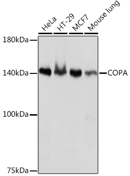 | Western blot analysis of various lysates using COPA Rabbit mAb (CAB19651) at 1:1000 dilution. Secondary antibody: HRP-conjugated Goat anti-Rabbit IgG (H+L) (CABS014) at 1:10000 dilution. Lysates/proteins: 25μg per lane. Blocking buffer: 3% nonfat dry milk in TBST. Detection: ECL Basic Kit (AbGn00020). Exposure time: 1s. |
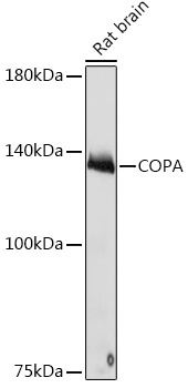 | Western blot analysis of lysates from Rat brain, using COPA Rabbit mAb (CAB19651) at 1:1000 dilution. Secondary antibody: HRP-conjugated Goat anti-Rabbit IgG (H+L) (CABS014) at 1:10000 dilution. Lysates/proteins: 25μg per lane. Blocking buffer: 3% nonfat dry milk in TBST. Detection: ECL Basic Kit (AbGn00020). Exposure time: 10s. |
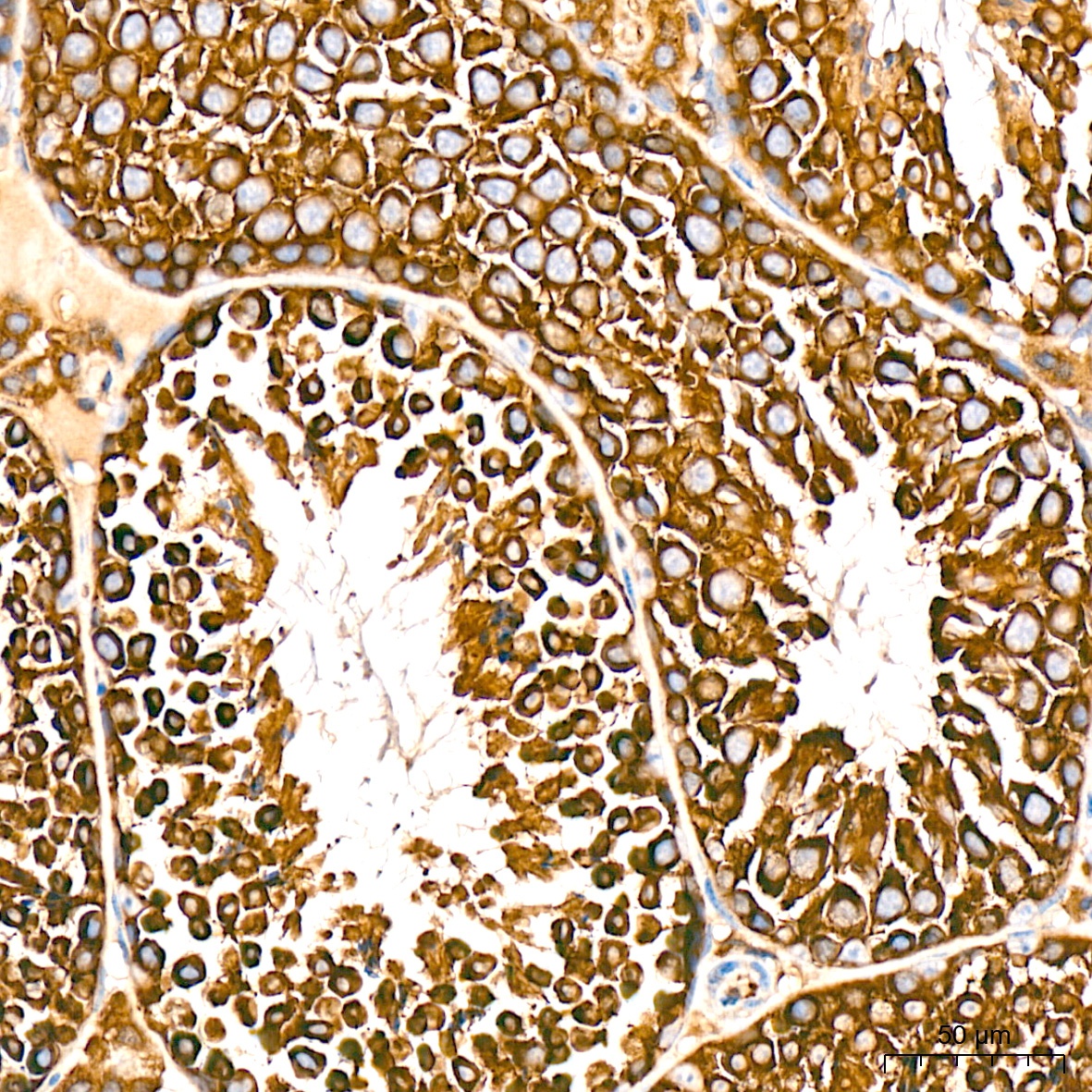 | Immunohistochemistry analysis of paraffin-embedded Mouse testis tissue using COPA Rabbit mAb (CAB19651) at a dilution of 1:200 (40x lens). High pressure antigen retrieval was performed with 0.01 M citrate buffer (pH 6.0) prior to IHC staining. |
 | Immunohistochemistry analysis of paraffin-embedded Human thyroid cancer tissue using COPA Rabbit mAb (CAB19651) at a dilution of 1:200 (40x lens). High pressure antigen retrieval was performed with 0.01 M citrate buffer (pH 6.0) prior to IHC staining. |
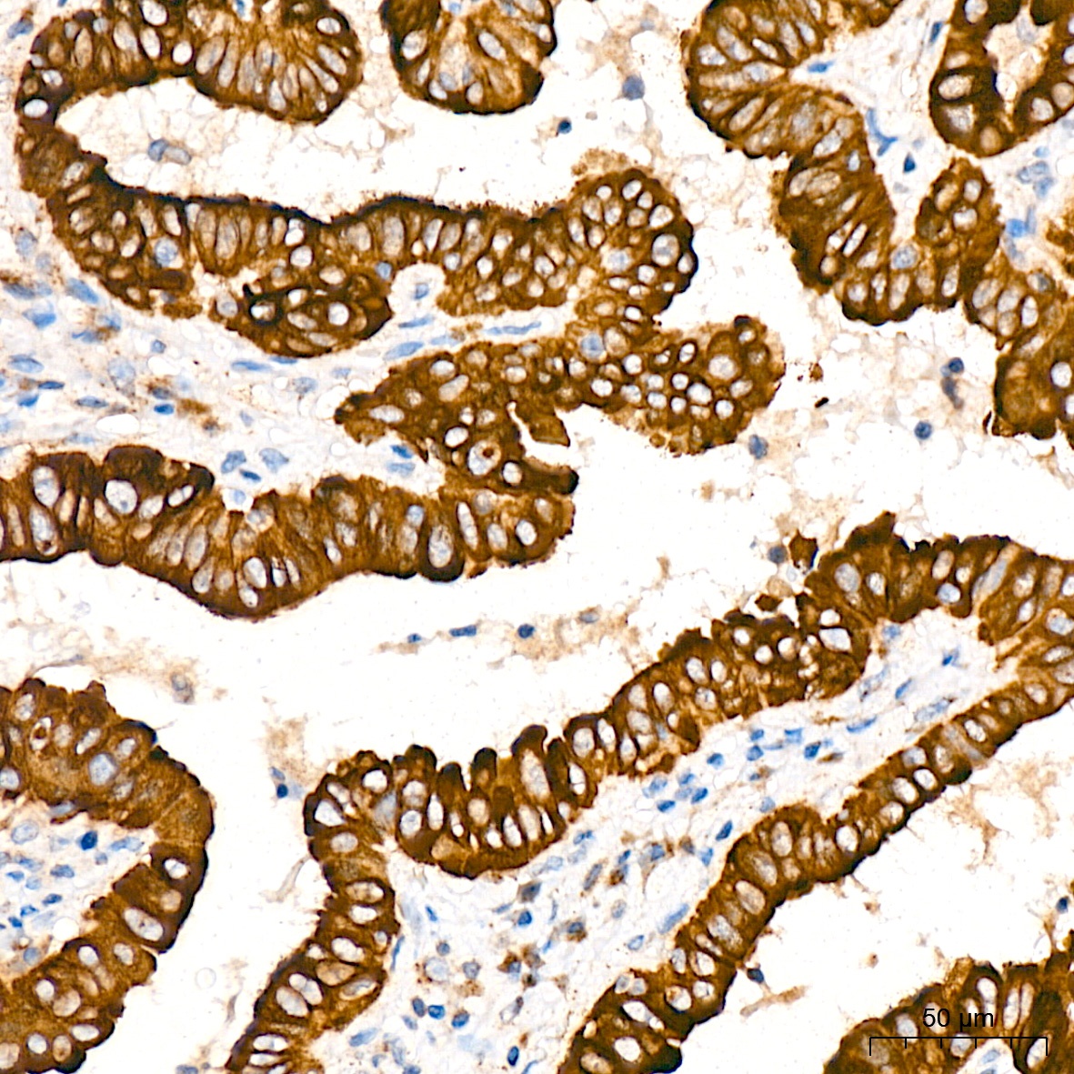
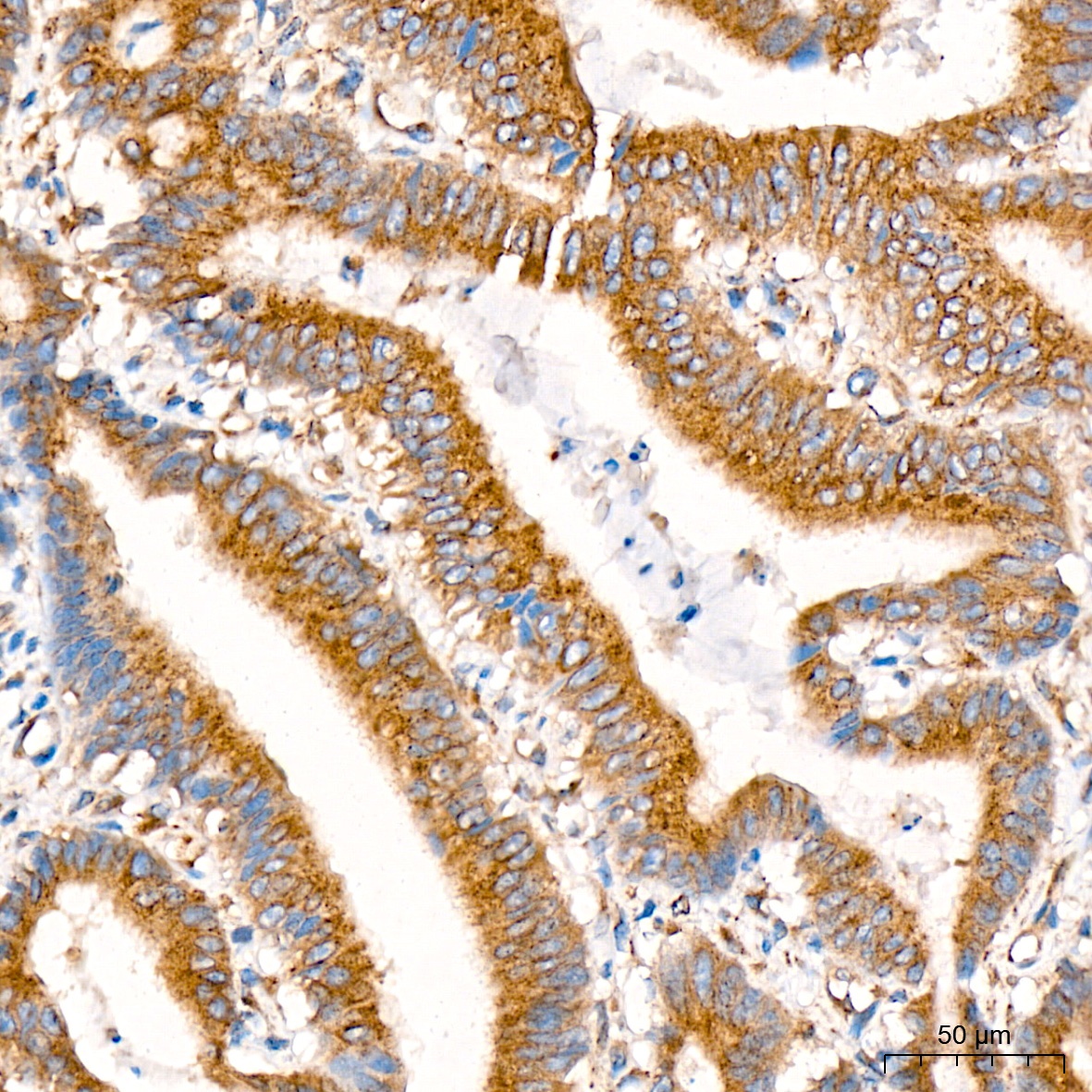 | Immunohistochemistry analysis of paraffin-embedded Human colon carcinoma tissue using COPA Rabbit mAb (CAB19651) at a dilution of 1:200 (40x lens). High pressure antigen retrieval was performed with 0.01 M citrate buffer (pH 6.0) prior to IHC staining. |
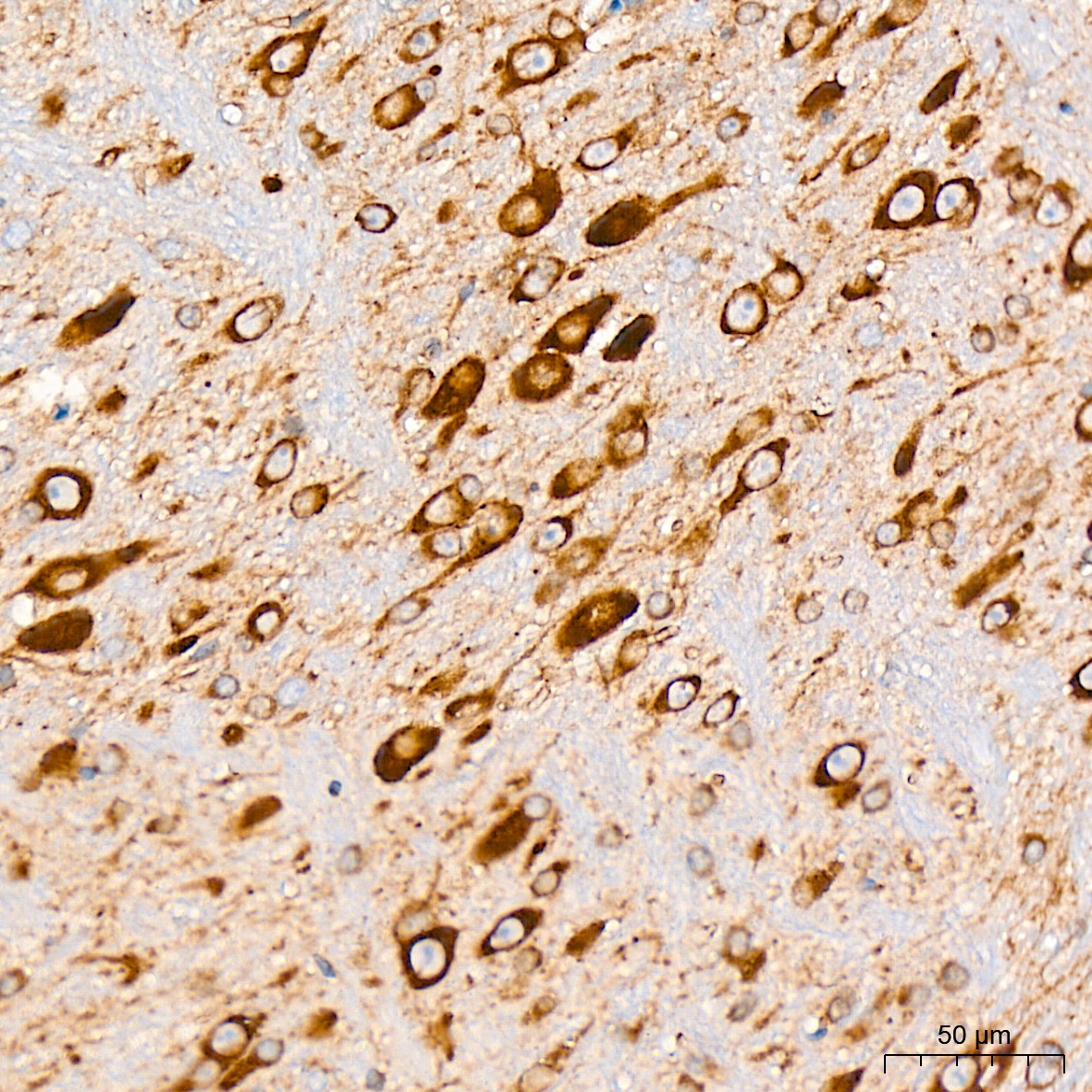 | Immunohistochemistry analysis of paraffin-embedded Mouse brain tissue using COPA Rabbit mAb (CAB19651) at a dilution of 1:200 (40x lens). High pressure antigen retrieval was performed with 0.01 M citrate buffer (pH 6.0) prior to IHC staining. |


