The DCR2 Monoclonal Antibody (CAB3581) is a high-quality antibody developed for reliable detection and analysis of target proteins. This antibody, generated in rabbits, has high reactivity with human samples, making it suitable for various applications, including Western blotting. It specifically targets the DCR2 protein, allowing for precise detection and analysis in different cell types.DCR2, also known as Decoy receptor 2, plays a crucial role in regulating apoptotic cell death and immune responses.
This antibody is validated for use in WB, IHC-P, ELISA applications and has demonstrated reactivity against Human, Mouse, Rat samples.
Product Name:
DCR2 Monoclonal Antibody
SKU:
CAB3581
Size:
20μL, 100μL
Reactivity:
Human, Mouse, Rat
Clone Number:
ARC2052
Conjugate:
Unconjugated
Immunogen:
Synthetic peptide. This information is considered to be commercially sensitive.
Recommended starting concentration is 1 μg/mL. Please optimize the concentration based on your specific assay requirements.
Synonyms:
DCR2, CD264, TRUNDD, TRAILR4, TRAIL-R4
Positive Sample:
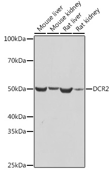
Mouse liver, Mouse kidney, Rat liver, Rat kidney
Cellular Localization:
Cell Surface, Plasma Membrane.
Calculated MW:
42kDa
Observed MW:
50kDa
The protein encoded by this gene is a member of the TNF-receptor superfamily. This receptor contains an extracellular TRAIL-binding domain, a transmembrane domain, and a truncated cytoplamic death domain. This receptor does not induce apoptosis, and has been shown to play an inhibitory role in TRAIL-induced cell apoptosis.
Purification Method
Affinity purification
Gene ID
8793
Buffer Information
Store at -20℃. Avoid freeze / thaw cycles. Buffer: PBS containing 50% glycerol and 0.05% BSA, preserved with proclin300 or sodium azide, pH 7.3.
Western blot analysis of various lysates using DCR2 Rabbit mAb (CAB3581) at 1:1000 dilution. Secondary antibody: HRP-conjugated Goat anti-Rabbit IgG (H+L) (CABS014) at 1:10000 dilution. Lysates/proteins: 25μg per lane. Blocking buffer: 3% nonfat dry milk in TBST. Detection: ECL Basic Kit (AbGn00020). Exposure time: 10s.
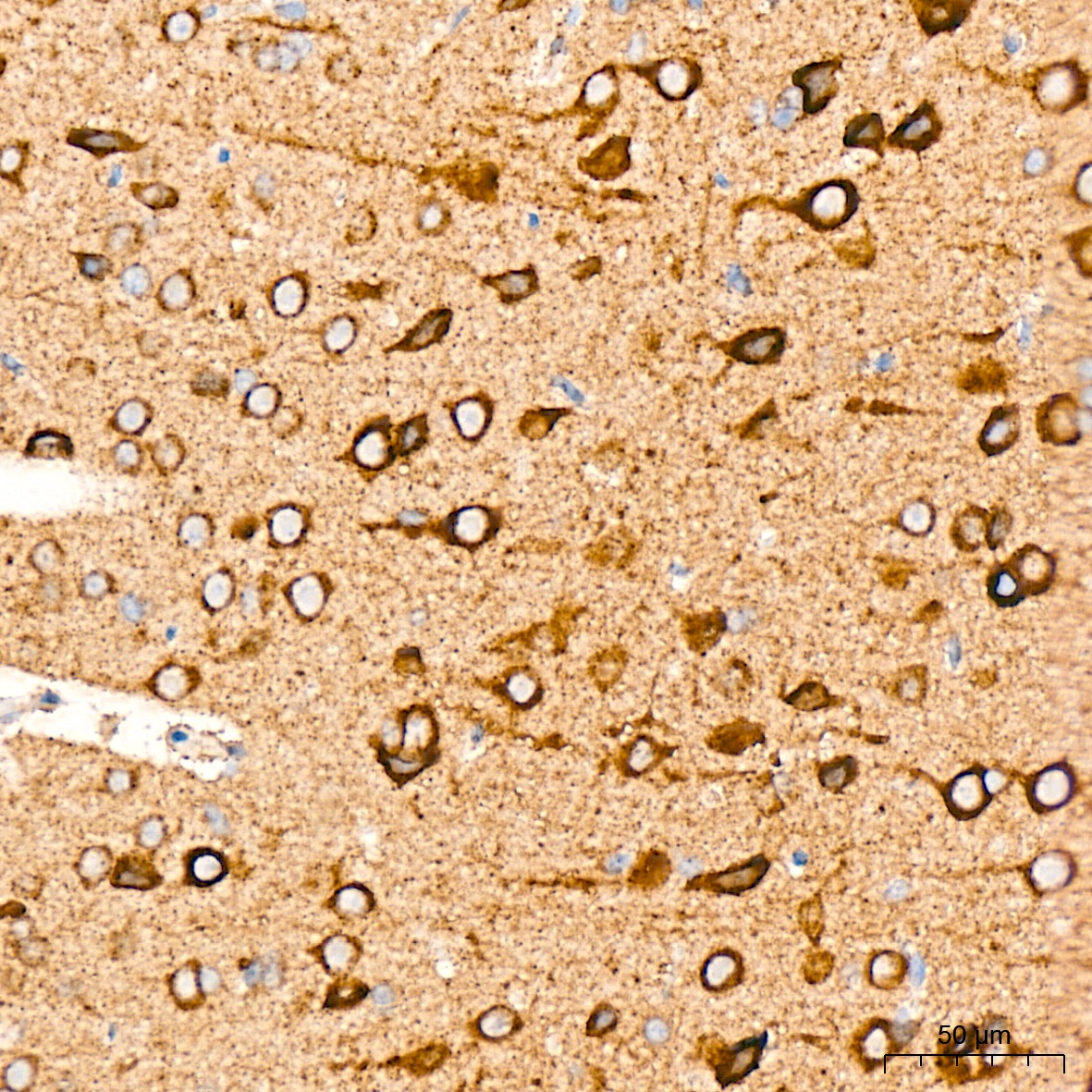
Immunohistochemistry analysis of paraffin-embedded Rat brain tissue using DCR2 Rabbit mAb (CAB3581) at a dilution of 1:200 (40x lens). High pressure antigen retrieval was performed with 0.01 M citrate buffer (pH 6.0) prior to IHC staining.
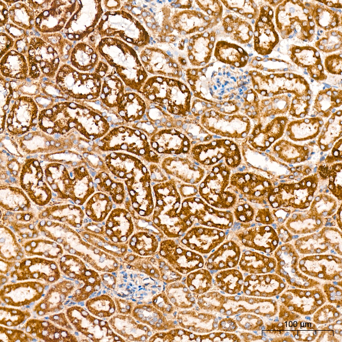
Immunohistochemistry analysis of paraffin-embedded Mouse kidney tissue using DCR2 Rabbit mAb (CAB3581) at a dilution of 1:200 (40x lens). High pressure antigen retrieval was performed with 0.01 M citrate buffer (pH 6.0) prior to IHC staining.
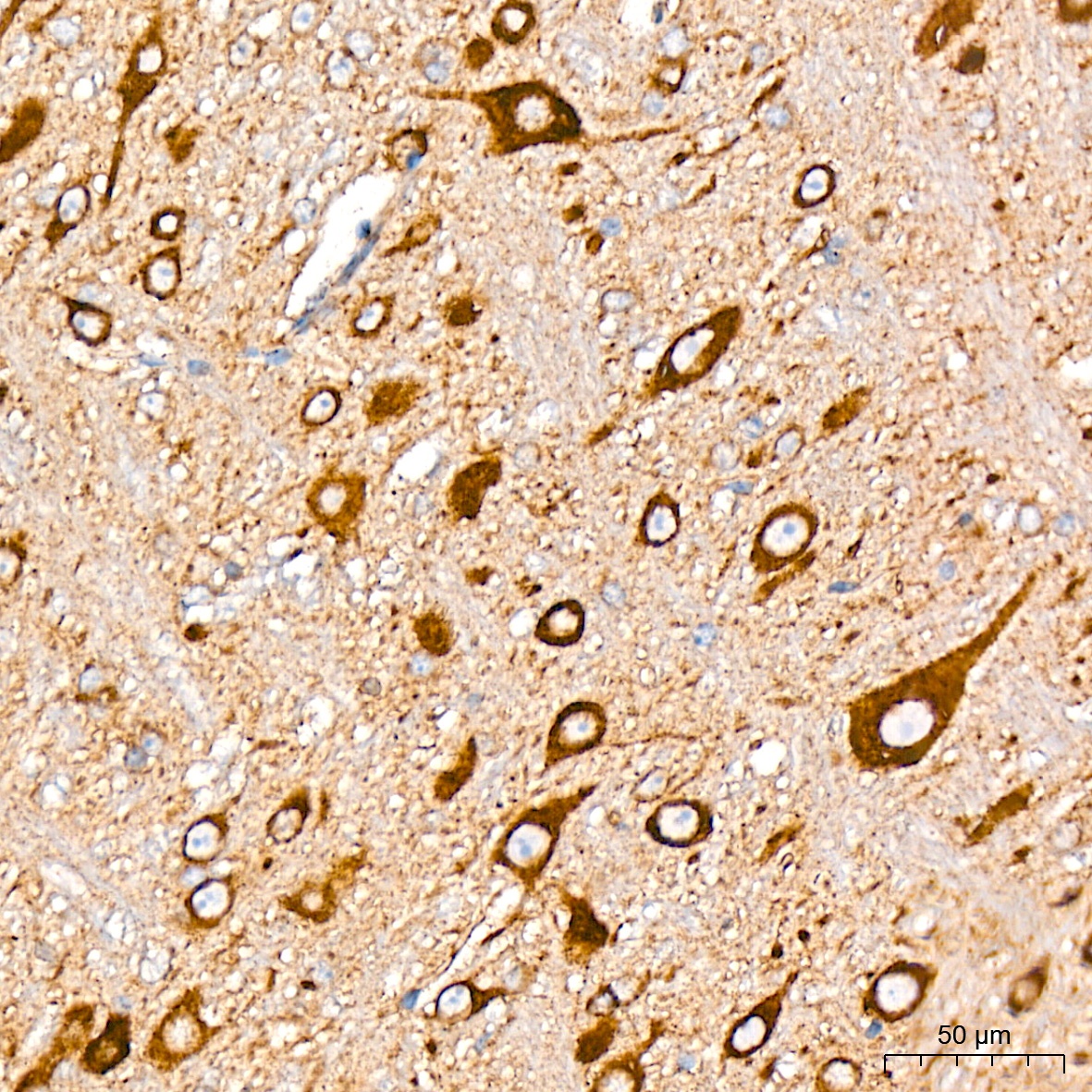
Immunohistochemistry analysis of paraffin-embedded Mouse brain tissue using DCR2 Rabbit mAb (CAB3581) at a dilution of 1:200 (40x lens). High pressure antigen retrieval was performed with 0.01 M citrate buffer (pH 6.0) prior to IHC staining.