The FADD Monoclonal Antibody (CAB19838) is a high-quality antibody developed for reliable detection and analysis of target proteins. This antibody, raised in rabbits, demonstrates high reactivity with human samples and is validated for use in Western blot applications. By binding specifically to the FADD protein, this antibody enables precise detection and analysis in a variety of cell types, making it ideal for investigations in immunology and cancer research.
This antibody is validated for use in WB, IHC-P, ELISA applications and has demonstrated reactivity against Human, Mouse, Rat samples.
Product Name:
FADD Monoclonal Antibody
SKU:
CAB19838
Size:
20μL, 100μL
Reactivity:
Human, Mouse, Rat
Clone Number:
ARC51937
Conjugate:
Unconjugated
Immunogen:
Recombinant protein (or fragment).This information is considered to be commercially sensitive.
Predicted to enable several functions, including caspase binding activity; death effector domain binding activity; and tumor necrosis factor receptor superfamily binding activity. Involved in several processes, including hematopoietic or lymphoid organ development; negative regulation of activation-induced cell death of T cells; and positive regulation of CD8-positive, alpha-beta cytotoxic T cell extravasation. Acts upstream of or within extrinsic apoptotic signaling pathway in absence of ligand; motor neuron apoptotic process; and regulation of programmed cell death. Predicted to be located in several cellular components, including cell body; cytosol; and membrane raft. Predicted to be part of CD95 death-inducing signaling complex and ripoptosome. Predicted to be active in cytoplasm. Is expressed in several structures, including alimentary system; brain; genitourinary system; hemolymphoid system gland; and liver and biliary system. Human ortholog(s) of this gene implicated in leukemia. Orthologous to human FADD (Fas associated via death domain).
Purification Method
Affinity purification
Gene ID
14082
Buffer Information
Store at -20℃. Avoid freeze / thaw cycles. Buffer: PBS containing 50% glycerol and 0.05% BSA, preserved with proclin300 or sodium azide, pH 7.3.
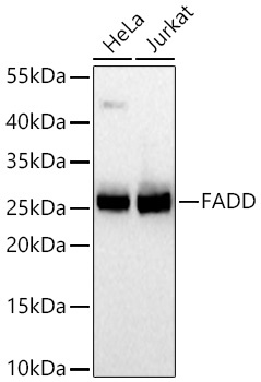
Western blot analysis of various lysates using FADD Rabbit mAb (CAB19838) at 1:1000 dilution incubated overnight at 4℃. Secondary antibody: HRP-conjugated Goat anti-Rabbit IgG (H+L) (CABS014) at 1:10000 dilution. Lysates/proteins: 25 μg per lane. Blocking buffer: 3% nonfat dry milk in TBST. Detection: ECL Basic Kit (AbGn00020). Exposure time: 90 s.
Western blot analysis of various lysates using FADD Rabbit mAb (CAB19838) at 1:1000 dilution incubated overnight at 4℃. Secondary antibody: HRP-conjugated Goat anti-Rabbit IgG (H+L) (CABS014) at 1:10000 dilution. Lysates/proteins: 25 μg per lane. Blocking buffer: 3% nonfat dry milk in TBST. Detection: ECL Basic Kit (AbGn00020). Exposure time: 90 s.
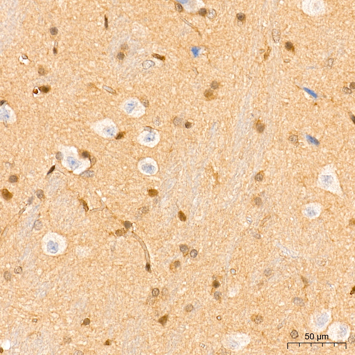
Immunohistochemistry analysis of paraffin-embedded Rat brain tissue using FADD Rabbit mAb (CAB19838) at a dilution of 1:200 (40x lens). High pressure antigen retrieval performed with 0.01M Tris-EDTA Buffer (pH 9.0) prior to IHC staining.
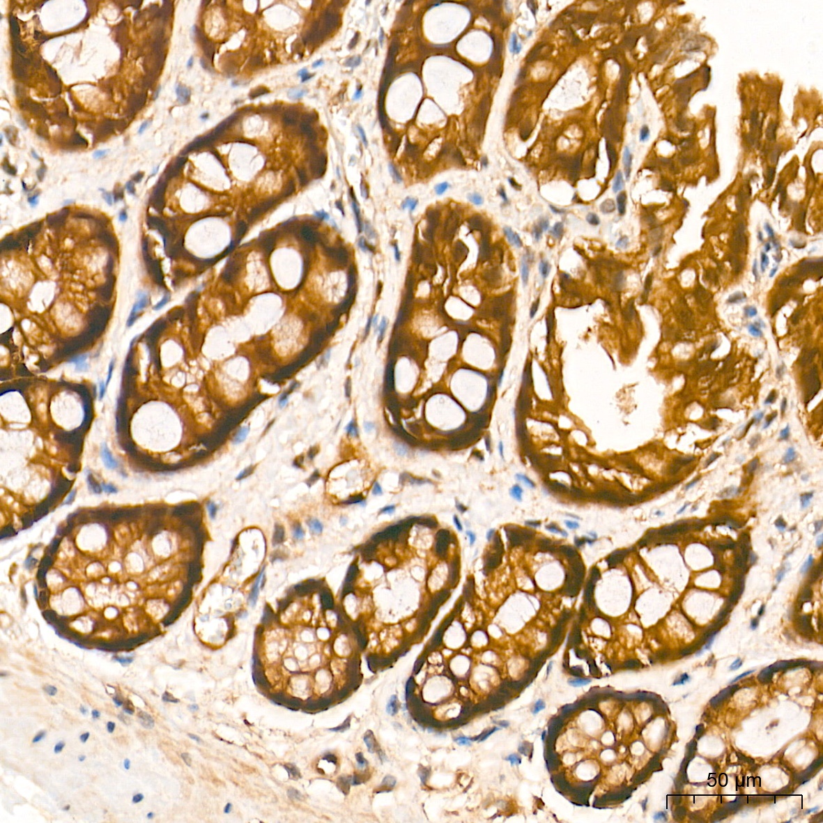
Immunohistochemistry analysis of paraffin-embedded Rat colon tissue using FADD Rabbit mAb (CAB19838) at a dilution of 1:200 (40x lens). High pressure antigen retrieval performed with 0.01M Tris-EDTA Buffer (pH 9.0) prior to IHC staining.